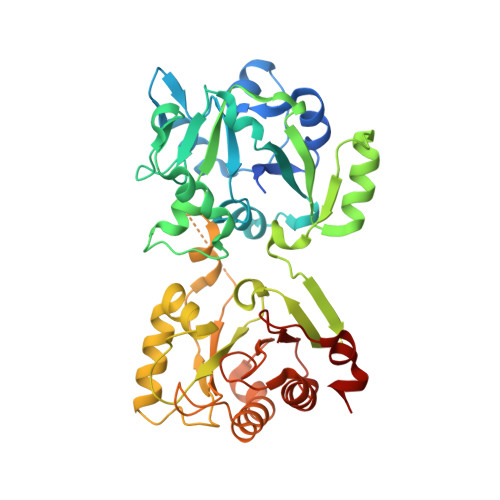
Molecular basis for DarT ADP-ribosylation of a DNA base.
Schuller, M., Butler, R.E., Ariza, A., Tromans-Coia, C., Jankevicius, G., Claridge, T.D.W., Kendall, S.L., Goh, S., Stewart, G.R., Ahel, I.(2021) Nature 596: 597-602
- PubMed: 34408320
- DOI: https://doi.org/10.1038/s41586-021-03825-4
- Primary Citation of Related Structures:
7OMU, 7OMV, 7OMW, 7OMX, 7OMY, 7OMZ, 7ON0 - PubMed Abstract:
ADP-ribosyltransferases use NAD + to catalyse substrate ADP-ribosylation 1 , and thereby regulate cellular pathways or contribute to toxin-mediated pathogenicity of bacteria 2-4 . Reversible ADP-ribosylation has traditionally been considered a protein-specific modification 5 , but recent in vitro studies have suggested nucleic acids as targets 6-9 . Here we present evidence that specific, reversible ADP-ribosylation of DNA on thymidine bases occurs in cellulo through the DarT-DarG toxin-antitoxin system, which is found in a variety of bacteria (including global pathogens such as Mycobacterium tuberculosis, enteropathogenic Escherichia coli and Pseudomonas aeruginosa) 10 . We report the structure of DarT, which identifies this protein as a diverged member of the PARP family. We provide a set of high-resolution structures of this enzyme in ligand-free and pre- and post-reaction states, which reveals a specialized mechanism of catalysis that includes a key active-site arginine that extends the canonical ADP-ribosyltransferase toolkit. Comparison with PARP-HPF1, a well-established DNA repair protein ADP-ribosylation complex, offers insights into how the DarT class of ADP-ribosyltransferases evolved into specific DNA-modifying enzymes. Together, our structural and mechanistic data provide details of this PARP family member and contribute to a fundamental understanding of the ADP-ribosylation of nucleic acids. We also show that thymine-linked ADP-ribose DNA adducts reversed by DarG antitoxin (functioning as a noncanonical DNA repair factor) are used not only for targeted DNA damage to induce toxicity, but also as a signalling strategy for cellular processes. Using M. tuberculosis as an exemplar, we show that DarT-DarG regulates growth by ADP-ribosylation of DNA at the origin of chromosome replication.
Organizational Affiliation:
Sir William Dunn School of Pathology, University of Oxford, Oxford, UK.


