7P3V

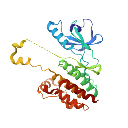
B-Raf V600E structure bound to a new inhibitor


Experimental Data Snapshot

  • Method: X-RAY DIFFRACTION
  • Resolution: 2.37 Å
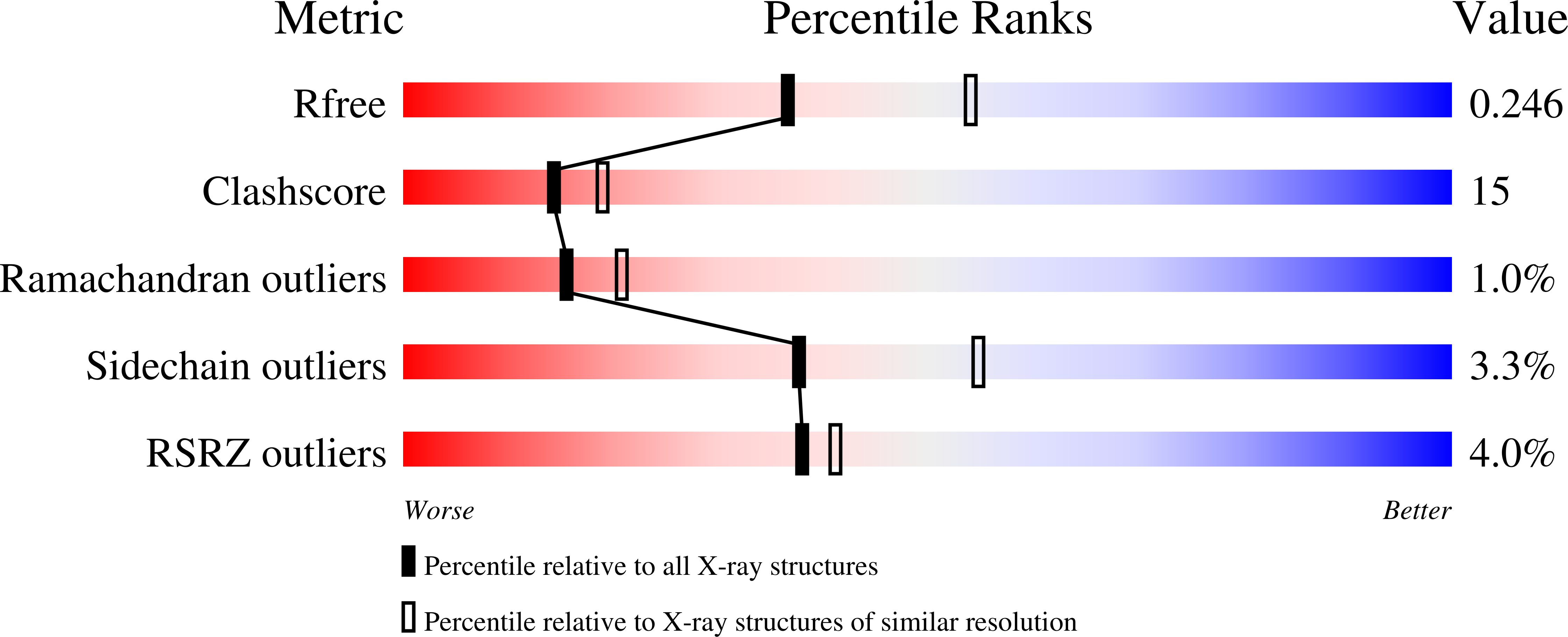
  • R-Value Free: 0.247 
  • R-Value Work: 0.195 
  • R-Value Observed: 0.199 

Starting Model: experimental
View more details

wwPDB Validation   3D Report Full Report


Ligand Structure Quality Assessment 


This is version 1.3 of the entry. See complete history


Literature

Structure-Based and Knowledge-Informed Design of B-Raf Inhibitors Devoid of Deleterious PXR Binding.

Schneider, M.Delfosse, V.Gelin, M.Grimaldi, M.Granell, M.Heriaud, L.Pons, J.L.Cohen Gonsaud, M.Balaguer, P.Bourguet, W.Labesse, G.

(2022) J Med Chem 65: 1552-1566

  • DOI: https://doi.org/10.1021/acs.jmedchem.1c01354
  • Primary Citation of Related Structures:  
    6HJ2, 7P3V

  • PubMed Abstract: 

    Dabrafenib is an anticancer drug currently used in the clinics, alone or in combination. However, dabrafenib was recently shown to potently activate the human nuclear receptor pregnane X receptor (PXR). PXR activation increases the clearance of various chemicals and drugs, including dabrafenib itself. It may also enhance cell proliferation and tumor aggressiveness. Therefore, there is a need for rational design of a potent protein kinase B-Raf inhibitor devoid of binding to the secondary target PXR and resisting rapid metabolism. By determining the crystal structure of dabrafenib bound to PXR and analyzing its mode of binding to both PXR and its primary target, B-Raf-V600E, we were able to derive new compounds with nanomolar activity against B-Raf and no detectable affinity for PXR. The crystal structure of B-Raf in complex with our lead compound revealed a subdomain swapping of the activation loop with potentially important functional implications for a prolonged inhibition of B-Raf-V600E.


  • Organizational Affiliation

    Centre de Biologie Structurale (CBS), CNRS, INSERM, Univ Montpellier, F-34090 Montpellier, France.


Macromolecules
Find similar proteins by:  (by identity cutoff)  |  3D Structure
Entity ID: 1
MoleculeChains Sequence LengthOrganismDetailsImage
Serine/threonine-protein kinase B-raf
A, B
275Homo sapiensMutation(s): 1 
Gene Names: BRAFBRAF1RAFB1
EC: 2.7.11.1
UniProt & NIH Common Fund Data Resources
Find proteins for P15056 (Homo sapiens)
Explore P15056 
Go to UniProtKB:  P15056
PHAROS:  P15056
GTEx:  ENSG00000157764 
Entity Groups  
Sequence Clusters30% Identity50% Identity70% Identity90% Identity95% Identity100% Identity
UniProt GroupP15056
Sequence Annotations
Expand
  • Reference Sequence
Small Molecules
Ligands 1 Unique
IDChains Name / Formula / InChI Key2D Diagram3D Interactions
5I4
Query on 5I4

Download Ideal Coordinates CCD File 
C [auth A],
D [auth B]
~{N}-[3-[5-(2-azanylpyrimidin-4-yl)-2-[(3~{S})-morpholin-3-yl]-1,3-thiazol-4-yl]-2-fluoranyl-phenyl]-2,5-bis(fluoranyl)benzenesulfonamide
C23 H19 F3 N6 O3 S2
SQCVSCOLOVRLEJ-KRWDZBQOSA-N
Experimental Data & Validation

Experimental Data

  • Method: X-RAY DIFFRACTION
  • Resolution: 2.37 Å
  • R-Value Free: 0.247 
  • R-Value Work: 0.195 
  • R-Value Observed: 0.199 
  • Space Group: P 21 21 21
Unit Cell:
Length ( Å )Angle ( ˚ )
a = 52.632α = 90
b = 104.854β = 90
c = 109.557γ = 90
Software Package:
Software NamePurpose
PHENIXrefinement
PHENIXrefinement
XDSdata reduction
XSCALEdata scaling
PHASERphasing

Structure Validation

View Full Validation Report


Ligand Structure Quality Assessment 


Entry History & Funding Information

Deposition Data


Funding OrganizationLocationGrant Number
French Infrastructure for Integrated Structural Biology (FRISBI)FranceANR-10-INBS-0005

Revision History  (Full details and data files)

  • Version 1.0: 2021-11-03
    Type: Initial release
  • Version 1.1: 2022-02-02
    Changes: Database references, Refinement description
  • Version 1.2: 2022-02-09
    Changes: Database references
  • Version 1.3: 2024-01-31
    Changes: Data collection, Refinement description