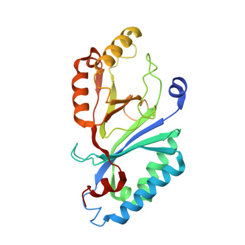
7Q4F

Structure of coproheme decarboxylase from Corynebacterium dipththeriae W183Y mutant in complex with coproheme


Experimental Data Snapshot

  • Method: X-RAY DIFFRACTION
  • Resolution: 2.15 Å
  • R-Value Free: 0.182 
  • R-Value Work: 0.140 
  • R-Value Observed: 0.142 

Starting Model: experimental
View more details

wwPDB Validation   3D Report Full Report


Ligand Structure Quality Assessment 


This is version 1.1 of the entry. See complete history


Literature

Initial Steps to Engineer Coproheme Decarboxylase to Obtain Stereospecific Monovinyl, Monopropionyl Deuterohemes.

Michlits, H.Valente, N.Mlynek, G.Hofbauer, S.

(2021) Front Bioeng Biotechnol 9: 807678-807678

  • DOI: https://doi.org/10.3389/fbioe.2021.807678
  • Primary Citation of Related Structures:  
    7Q4F, 7Q4G

  • PubMed Abstract: 

    The oxidative decarboxylation of coproheme to form heme b by coproheme decarboxylase is a stereospecific two-step reaction. In the first step, the propionate at position two (p2) is cleaved off the pyrrole ring A to form a vinyl group at this position. Subsequently, the propionate at position four (p4) on pyrrole ring B is cleaved off and heme b is formed. In this study, we attempted to engineer coproheme decarboxylase from Corynebacterium diphtheriae to alter the stereospecificity of this reaction. By introducing a tyrosine residue in proximity to the propionate at position 4, we were able to create a new radical center in the active site. However, the artificial Tyr183 radical could not be shown to catalyze any decarboxylation.


  • Organizational Affiliation

    Department of Chemistry, Institute of Biochemistry, University of Natural Resources and Life Sciences, Vienna, Austria.


Macromolecules
Find similar proteins by:  (by identity cutoff)  |  3D Structure
Entity ID: 1
MoleculeChains Sequence LengthOrganismDetailsImage
Coproheme decarboxylase from Corynebacterium dipththeriae W183Y mutant in complex with coproheme
A, B, C, D, E
237Corynebacterium diphtheriaeMutation(s): 1 
Gene Names: BT093_04375
EC: 1.3.98.5
UniProt
Find proteins for Q6NGV6 (Corynebacterium diphtheriae (strain ATCC 700971 / NCTC 13129 / Biotype gravis))
Explore Q6NGV6 
Go to UniProtKB:  Q6NGV6
Entity Groups  
Sequence Clusters30% Identity50% Identity70% Identity90% Identity95% Identity100% Identity
UniProt GroupQ6NGV6
Sequence Annotations
Expand
  • Reference Sequence
Small Molecules
Ligands 3 Unique
IDChains Name / Formula / InChI Key2D Diagram3D Interactions
FEC (Subject of Investigation/LOI)
Query on FEC

Download Ideal Coordinates CCD File 
F [auth A],
H [auth B],
J [auth C],
K [auth D],
L [auth E]
1,3,5,8-TETRAMETHYL-PORPHINE-2,4,6,7-TETRAPROPIONIC ACID FERROUS COMPLEX
C36 H36 Fe N4 O8
FEDZMOFKVKOYTI-RGGAHWMASA-L
PGE
Query on PGE

Download Ideal Coordinates CCD File 
G [auth A]TRIETHYLENE GLYCOL
C6 H14 O4
ZIBGPFATKBEMQZ-UHFFFAOYSA-N
PEG
Query on PEG

Download Ideal Coordinates CCD File 
I [auth B],
M [auth E]
DI(HYDROXYETHYL)ETHER
C4 H10 O3
MTHSVFCYNBDYFN-UHFFFAOYSA-N
Experimental Data & Validation

Experimental Data

  • Method: X-RAY DIFFRACTION
  • Resolution: 2.15 Å
  • R-Value Free: 0.182 
  • R-Value Work: 0.140 
  • R-Value Observed: 0.142 
  • Space Group: P 3 1 2
Unit Cell:
Length ( Å )Angle ( ˚ )
a = 141.809α = 90
b = 141.809β = 90
c = 124.863γ = 120
Software Package:
Software NamePurpose
PDB-REDOrefinement
PHENIXrefinement
XDSdata reduction
EDNAdata collection
PHASERphasing

Structure Validation

View Full Validation Report


Ligand Structure Quality Assessment 


Entry History & Funding Information

Deposition Data


Funding OrganizationLocationGrant Number
Austrian Science FundAustriaP29099

Revision History  (Full details and data files)

  • Version 1.0: 2022-02-23
    Type: Initial release
  • Version 1.1: 2024-01-31
    Changes: Data collection, Refinement description