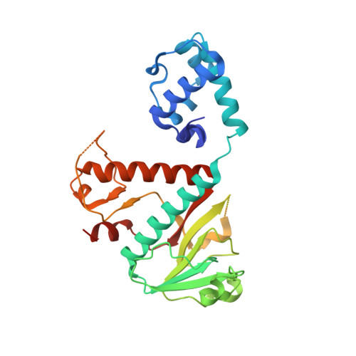
7TW9

Structure of nsp14 N7-MethylTransferase domain fused with TELSAM bound to Sinefungin


Experimental Data Snapshot

  • Method: X-RAY DIFFRACTION
  • Resolution: 1.41 Å
  • R-Value Free: 0.232 
  • R-Value Work: 0.192 
  • R-Value Observed: 0.194 

Starting Models: experimental
View more details

wwPDB Validation   3D Report Full Report


Ligand Structure Quality Assessment 


This is version 1.3 of the entry. See complete history


Literature

High-resolution structures of the SARS-CoV-2 N7-methyltransferase inform therapeutic development.

Kottur, J.Rechkoblit, O.Quintana-Feliciano, R.Sciaky, D.Aggarwal, A.K.

(2022) Nat Struct Mol Biol 29: 850-853

  • DOI: https://doi.org/10.1038/s41594-022-00828-1
  • Primary Citation of Related Structures:  
    7TW7, 7TW8, 7TW9

  • PubMed Abstract: 

    Emergence of SARS-CoV-2 coronavirus has led to millions of deaths globally. We present three high-resolution crystal structures of the SARS-CoV-2 nsp14 N7-methyltransferase core bound to S-adenosylmethionine (1.62 Å), S-adenosylhomocysteine (1.55 Å) and sinefungin (1.41 Å). We identify features of the methyltransferase core that are crucial for the development of antivirals and show SAH as the best scaffold for the design of antivirals against SARS-CoV-2 and other pathogenic coronaviruses.


  • Organizational Affiliation

    Department of Pharmacological Sciences, Icahn School of Medicine at Mount Sinai, New York, NY, USA. [email protected].


Macromolecules
Find similar proteins by:  (by identity cutoff)  |  3D Structure
Entity ID: 1
MoleculeChains Sequence LengthOrganismDetailsImage
Transcription factor ETV6,Proofreading exoribonuclease nsp14 chimera309Severe acute respiratory syndrome coronavirus 2Mutation(s): 3 
Gene Names: ETV6TELTEL1rep1a-1b
EC: 3.1.13
UniProt & NIH Common Fund Data Resources
Find proteins for P41212 (Homo sapiens)
Explore P41212 
Go to UniProtKB:  P41212
PHAROS:  P41212
GTEx:  ENSG00000139083 
Find proteins for P0DTD1 (Severe acute respiratory syndrome coronavirus 2)
Explore P0DTD1 
Go to UniProtKB:  P0DTD1
Entity Groups  
Sequence Clusters30% Identity50% Identity70% Identity90% Identity95% Identity100% Identity
UniProt GroupsP0DTD1P41212
Sequence Annotations
Expand
  • Reference Sequence
Small Molecules
Ligands 5 Unique
IDChains Name / Formula / InChI Key2D Diagram3D Interactions
SFG (Subject of Investigation/LOI)
Query on SFG

Download Ideal Coordinates CCD File 
B [auth A]SINEFUNGIN
C15 H23 N7 O5
LMXOHSDXUQEUSF-YECHIGJVSA-N
PEG
Query on PEG

Download Ideal Coordinates CCD File 
D [auth A]DI(HYDROXYETHYL)ETHER
C4 H10 O3
MTHSVFCYNBDYFN-UHFFFAOYSA-N
ZN
Query on ZN

Download Ideal Coordinates CCD File 
C [auth A]ZINC ION
Zn
PTFCDOFLOPIGGS-UHFFFAOYSA-N
EOH
Query on EOH

Download Ideal Coordinates CCD File 
E [auth A]ETHANOL
C2 H6 O
LFQSCWFLJHTTHZ-UHFFFAOYSA-N
MOH
Query on MOH

Download Ideal Coordinates CCD File 
F [auth A],
G [auth A],
H [auth A],
I [auth A],
J [auth A]
METHANOL
C H4 O
OKKJLVBELUTLKV-UHFFFAOYSA-N
Experimental Data & Validation

Experimental Data

  • Method: X-RAY DIFFRACTION
  • Resolution: 1.41 Å
  • R-Value Free: 0.232 
  • R-Value Work: 0.192 
  • R-Value Observed: 0.194 
  • Space Group: P 65
Unit Cell:
Length ( Å )Angle ( ˚ )
a = 109.028α = 90
b = 109.028β = 90
c = 48.641γ = 120
Software Package:
Software NamePurpose
Aimlessdata scaling
PHENIXrefinement
PDB_EXTRACTdata extraction
DIALSdata reduction
STARANISOdata scaling
PHASERphasing

Structure Validation

View Full Validation Report


Ligand Structure Quality Assessment 


Entry History & Funding Information

Deposition Data


Funding OrganizationLocationGrant Number
Not funded--

Revision History  (Full details and data files)

  • Version 1.0: 2022-09-07
    Type: Initial release
  • Version 1.1: 2022-09-21
    Changes: Database references
  • Version 1.2: 2022-10-05
    Changes: Database references
  • Version 1.3: 2023-10-18
    Changes: Data collection, Refinement description