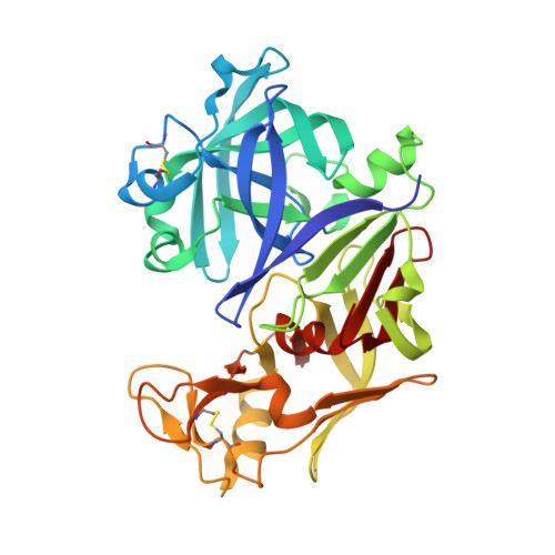
7VE0

Crystal Structure of Ritonavir bound Plasmepsin II (PMII) from Plasmodium falciparum


Experimental Data Snapshot

  • Method: X-RAY DIFFRACTION
  • Resolution: 1.90 Å
  • R-Value Free: 0.261 
  • R-Value Work: 0.222 
  • R-Value Observed: 0.224 

Starting Model: experimental
View more details

wwPDB Validation   3D Report Full Report


Ligand Structure Quality Assessment 


This is version 1.3 of the entry. See complete history


Literature

Inhibition of Plasmodium falciparum plasmepsins by drugs targeting HIV-1 protease: A way forward for antimalarial drug discovery.

Mishra, V.Deshmukh, A.Rathore, I.Chakraborty, S.Patankar, S.Gustchina, A.Wlodawer, A.Yada, R.Y.Bhaumik, P.

(2024) Curr Res Struct Biol 7: 100128-100128

  • DOI: https://doi.org/10.1016/j.crstbi.2024.100128
  • Primary Citation of Related Structures:  
    7VE0, 7VE2

  • PubMed Abstract: 

    Plasmodium species are causative agents of malaria, a disease that is a serious global health concern. FDA-approved HIV-1 protease inhibitors (HIV-1 PIs) have been reported to be effective in reducing the infection by Plasmodium parasites in the population co-infected with both HIV-1 and malaria. However, the mechanism of HIV-1 PIs in mitigating Plasmodium pathogenesis during malaria/HIV-1 co-infection is not fully understood. In this study we demonstrate that HIV-1 drugs ritonavir (RTV) and lopinavir (LPV) exhibit the highest inhibition activity against plasmepsin II (PMII) and plasmepsin X (PMX) of P. falciparum. Crystal structures of the complexes of PMII with both drugs have been determined. The inhibitors interact with PMII via multiple hydrogen bonding and hydrophobic interactions. The P4 moiety of RTV forms additional interactions compared to LPV and exhibits conformational flexibility in a large S4 pocket of PMII. Our study is also the first to report inhibition of P. falciparum PMX by RTV and the mode of binding of the drug to the PMX active site. Analysis of the crystal structures implies that PMs can accommodate bulkier groups of these inhibitors in their S4 binding pockets. Structurally similar active sites of different vacuolar and non-vacuolar PMs suggest the potential of HIV-1 PIs in targeting these enzymes with differential affinities. Our structural investigations and biochemical data emphasize PMs as crucial targets for repurposing HIV-1 PIs as antimalarial drugs.


  • Organizational Affiliation

    Department of Biosciences and Bioengineering, Indian Institute of Technology Bombay, Powai, Mumbai, 400076, India.


Macromolecules
Find similar proteins by:  (by identity cutoff)  |  3D Structure
Entity ID: 1
MoleculeChains Sequence LengthOrganismDetailsImage
Plasmepsin II
A, B
331Plasmodium falciparum 3D7Mutation(s): 0 
Gene Names: PF3D7_1408000
EC: 3.4.23.39
UniProt
Find proteins for Q8I6V3 (Plasmodium falciparum (isolate 3D7))
Explore Q8I6V3 
Go to UniProtKB:  Q8I6V3
Entity Groups  
Sequence Clusters30% Identity50% Identity70% Identity90% Identity95% Identity100% Identity
UniProt GroupQ8I6V3
Sequence Annotations
Expand
  • Reference Sequence
Small Molecules
Ligands 3 Unique
IDChains Name / Formula / InChI Key2D Diagram3D Interactions
RIT (Subject of Investigation/LOI)
Query on RIT

Download Ideal Coordinates CCD File 
C [auth A],
L [auth B]
RITONAVIR
C37 H48 N6 O5 S2
NCDNCNXCDXHOMX-XGKFQTDJSA-N
CPS
Query on CPS

Download Ideal Coordinates CCD File 
D [auth A]
E [auth A]
F [auth A]
G [auth A]
H [auth A]
D [auth A],
E [auth A],
F [auth A],
G [auth A],
H [auth A],
I [auth A],
J [auth A],
M [auth B],
N [auth B],
O [auth B],
P [auth B],
Q [auth B],
R [auth B],
S [auth B],
T [auth B]
3-[(3-CHOLAMIDOPROPYL)DIMETHYLAMMONIO]-1-PROPANESULFONATE
C32 H58 N2 O7 S
UMCMPZBLKLEWAF-BCTGSCMUSA-N
EDO
Query on EDO

Download Ideal Coordinates CCD File 
K [auth A],
U [auth B]
1,2-ETHANEDIOL
C2 H6 O2
LYCAIKOWRPUZTN-UHFFFAOYSA-N
Biologically Interesting Molecules (External Reference) 1 Unique
Experimental Data & Validation

Experimental Data

  • Method: X-RAY DIFFRACTION
  • Resolution: 1.90 Å
  • R-Value Free: 0.261 
  • R-Value Work: 0.222 
  • R-Value Observed: 0.224 
  • Space Group: C 1 2 1
Unit Cell:
Length ( Å )Angle ( ˚ )
a = 150.29α = 90
b = 70.41β = 133.61
c = 106.67γ = 90
Software Package:
Software NamePurpose
REFMACrefinement
XDSdata reduction
XSCALEdata scaling
PHASERphasing

Structure Validation

View Full Validation Report


Ligand Structure Quality Assessment 


Entry History & Funding Information

Deposition Data


Funding OrganizationLocationGrant Number
Department of Biotechnology (DBT, India)IndiaRamalingaswami Re-entry Fellowship

Revision History  (Full details and data files)

  • Version 1.0: 2023-02-01
    Type: Initial release
  • Version 1.1: 2023-11-29
    Changes: Data collection, Refinement description
  • Version 1.2: 2024-03-06
    Changes: Database references
  • Version 1.3: 2024-10-23
    Changes: Structure summary