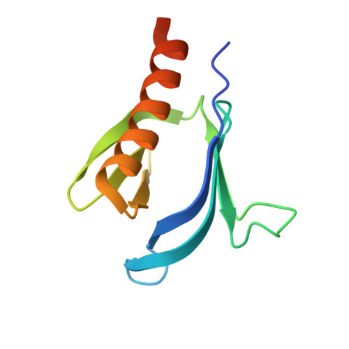
7YIR

Crystal structure of N-terminal PH domain of ARAP3 protein from human


Experimental Data Snapshot

  • Method: X-RAY DIFFRACTION
  • Resolution: 2.10 Å
  • R-Value Free: 0.230 
  • R-Value Work: 0.184 
  • R-Value Observed: 0.187 

Starting Model: experimental
View more details

wwPDB Validation   3D Report Full Report


Ligand Structure Quality Assessment 


This is version 1.1 of the entry. See complete history


Literature

Structural Insights Uncover the Specific Phosphoinositide Recognition by the PH1 Domain of Arap3.

Zhang, Y.Ge, L.Xu, L.Liu, Y.Wang, J.Liu, C.Zhao, H.Xing, L.Wang, J.Wu, B.

(2023) Int J Mol Sci 24

  • DOI: https://doi.org/10.3390/ijms24021125
  • Primary Citation of Related Structures:  
    7YIR, 7YIS

  • PubMed Abstract: 

    Arap3, a dual GTPase-activating protein (GAP) for the small GTPases Arf6 and RhoA, plays key roles in regulating a wide range of biological processes, including cancer cell invasion and metastasis. It is known that Arap3 is a PI3K effector that can bind directly to PI(3,4,5)P3, and the PI(3,4,5)P3-mediated plasma membrane recruitment is crucial for its function. However, the molecular mechanism of how the protein recognizes PI(3,4,5)P3 remains unclear. Here, using liposome pull-down and surface plasmon resonance (SPR) analysis, we found that the N-terminal first pleckstrin homology (PH) domain (Arap3-PH1) can interact with PI(3,4,5)P3 and, with lower affinity, with PI(4,5)P2. To understand how Arap3-PH1 and phosphoinositide (PIP) lipids interact, we solved the crystal structure of the Arap3-PH1 in the apo form and complex with diC4-PI(3,4,5)P3. We also characterized the interactions of Arap3-PH1 with diC4-PI(3,4,5)P3 and diC4-PI(4,5)P2 in solution by nuclear magnetic resonance (NMR) spectroscopy. Furthermore, we found overexpression of Arap3 could inhibit breast cancer cell invasion in vitro, and the PIPs-binding ability of the PH1 domain is essential for this function.


  • Organizational Affiliation

    High Magnetic Field Laboratory, Key Laboratory of High Magnetic Field and Ion Beam Physical Biology, Hefei Institutes of Physical Science, Chinese Academy of Sciences, Hefei 230031, China.


Macromolecules
Find similar proteins by:  (by identity cutoff)  |  3D Structure
Entity ID: 1
MoleculeChains Sequence LengthOrganismDetailsImage
Arf-GAP with Rho-GAP domain, ANK repeat and PH domain-containing protein 3111Homo sapiensMutation(s): 0 
Gene Names: ARAP3CENTD3
UniProt & NIH Common Fund Data Resources
Find proteins for Q8WWN8 (Homo sapiens)
Explore Q8WWN8 
Go to UniProtKB:  Q8WWN8
PHAROS:  Q8WWN8
GTEx:  ENSG00000120318 
Entity Groups  
Sequence Clusters30% Identity50% Identity70% Identity90% Identity95% Identity100% Identity
UniProt GroupQ8WWN8
Sequence Annotations
Expand
  • Reference Sequence
Small Molecules
Ligands 1 Unique
IDChains Name / Formula / InChI Key2D Diagram3D Interactions
PEG (Subject of Investigation/LOI)
Query on PEG

Download Ideal Coordinates CCD File 
B [auth A]DI(HYDROXYETHYL)ETHER
C4 H10 O3
MTHSVFCYNBDYFN-UHFFFAOYSA-N
Experimental Data & Validation

Experimental Data

  • Method: X-RAY DIFFRACTION
  • Resolution: 2.10 Å
  • R-Value Free: 0.230 
  • R-Value Work: 0.184 
  • R-Value Observed: 0.187 
  • Space Group: P 61 2 2
Unit Cell:
Length ( Å )Angle ( ˚ )
a = 102.634α = 90
b = 102.634β = 90
c = 45.506γ = 120
Software Package:
Software NamePurpose
SCALAdata scaling
PHENIXrefinement
PDB_EXTRACTdata extraction
HKL-2000data reduction
PHASERphasing

Structure Validation

View Full Validation Report


Ligand Structure Quality Assessment 


Entry History & Funding Information

Deposition Data


Funding OrganizationLocationGrant Number
Not funded--

Revision History  (Full details and data files)

  • Version 1.0: 2023-05-03
    Type: Initial release
  • Version 1.1: 2023-11-29
    Changes: Data collection, Refinement description