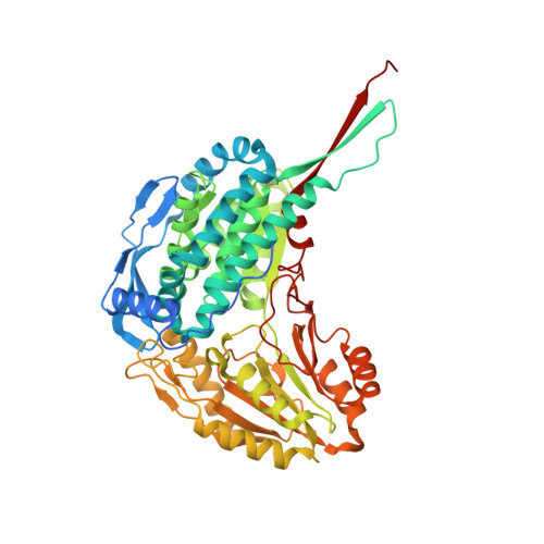
7MES

Structure of ALDH4A1 complexed with trans-4-Hydroxy-D-proline


Experimental Data Snapshot

  • Method: X-RAY DIFFRACTION
  • Resolution: 1.37 Å
  • R-Value Free: 0.172 
  • R-Value Work: 0.161 
  • R-Value Observed: 0.161 

Starting Model: experimental
View more details

wwPDB Validation   3D Report Full Report


Ligand Structure Quality Assessment 


This is version 1.2 of the entry. See complete history


Literature

Structural basis for the stereospecific inhibition of the dual proline/hydroxyproline catabolic enzyme ALDH4A1 by trans-4-hydroxy-L-proline.

Bogner, A.N.Stiers, K.M.McKay, C.M.Becker, D.F.Tanner, J.J.

(2021) Protein Sci 30: 1714-1722

  • DOI: https://doi.org/10.1002/pro.4131
  • Primary Citation of Related Structures:  
    7MER, 7MES

  • PubMed Abstract: 

    Aldehyde dehydrogenase 4A1 (ALDH4A1) catalyzes the final steps of both proline and hydroxyproline catabolism. It is a dual substrate enzyme that catalyzes the NAD + -dependent oxidations of L-glutamate-γ-semialdehyde to L-glutamate (proline metabolism), and 4-hydroxy-L-glutamate-γ-semialdehyde to 4-erythro-hydroxy-L-glutamate (hydroxyproline metabolism). Here we investigated the inhibition of mouse ALDH4A1 by the six stereoisomers of proline and 4-hydroxyproline using steady-state kinetics and X-ray crystallography. Trans-4-hydroxy-L-proline is the strongest of the inhibitors studied, characterized by a competitive inhibition constant of 0.7 mM, followed by L-proline (1.9 mM). The other compounds are very weak inhibitors (approximately 10 mM or greater). Insight into the selectivity for L-stereoisomers was obtained by solving crystal structures of ALDH4A1 complexed with trans-4-hydroxy-L-proline and trans-4-hydroxy-D-proline. The structures suggest that the 10-fold greater preference for the L-stereoisomer is due to a serine residue that hydrogen bonds to the amine group of trans-4-hydroxy-L-proline. In contrast, the amine group of the D-stereoisomer lacks a direct interaction with the enzyme due to a different orientation of the pyrrolidine ring. These results suggest that hydroxyproline catabolism is subject to substrate inhibition by trans-4-hydroxy-L-proline, analogous to the known inhibition of proline catabolism by L-proline. Also, drugs targeting the first enzyme of hydroxyproline catabolism, by elevating the level of trans-4-hydroxy-L-proline, may inadvertently impair proline catabolism by the inhibition of ALDH4A1.


  • Organizational Affiliation

    Department of Biochemistry, University of Missouri, Columbia, Missouri, USA.


Macromolecules
Find similar proteins by:  (by identity cutoff)  |  3D Structure
Entity ID: 1
MoleculeChains Sequence LengthOrganismDetailsImage
Delta-1-pyrroline-5-carboxylate dehydrogenase, mitochondrial
A, B
563Mus musculusMutation(s): 0 
Gene Names: Aldh4a1
EC: 1.2.1.88
UniProt & NIH Common Fund Data Resources
Find proteins for Q8CHT0 (Mus musculus)
Explore Q8CHT0 
Go to UniProtKB:  Q8CHT0
IMPC:  MGI:2443883
Entity Groups  
Sequence Clusters30% Identity50% Identity70% Identity90% Identity95% Identity100% Identity
UniProt GroupQ8CHT0
Sequence Annotations
Expand
  • Reference Sequence
Small Molecules
Ligands 6 Unique
IDChains Name / Formula / InChI Key2D Diagram3D Interactions
NAD (Subject of Investigation/LOI)
Query on NAD

Download Ideal Coordinates CCD File 
C [auth A],
J [auth B]
NICOTINAMIDE-ADENINE-DINUCLEOTIDE
C21 H27 N7 O14 P2
BAWFJGJZGIEFAR-NNYOXOHSSA-N
1PE
Query on 1PE

Download Ideal Coordinates CCD File 
I [auth A],
O [auth B]
PENTAETHYLENE GLYCOL
C10 H22 O6
JLFNLZLINWHATN-UHFFFAOYSA-N
PG4
Query on PG4

Download Ideal Coordinates CCD File 
F [auth A]TETRAETHYLENE GLYCOL
C8 H18 O5
UWHCKJMYHZGTIT-UHFFFAOYSA-N
PGE
Query on PGE

Download Ideal Coordinates CCD File 
M [auth B]TRIETHYLENE GLYCOL
C6 H14 O4
ZIBGPFATKBEMQZ-UHFFFAOYSA-N
UY7 (Subject of Investigation/LOI)
Query on UY7

Download Ideal Coordinates CCD File 
D [auth A],
E [auth A],
K [auth B],
L [auth B]
(4S)-4-hydroxy-D-proline
C5 H9 N O3
PMMYEEVYMWASQN-IUYQGCFVSA-N
PEG
Query on PEG

Download Ideal Coordinates CCD File 
G [auth A],
H [auth A],
N [auth B]
DI(HYDROXYETHYL)ETHER
C4 H10 O3
MTHSVFCYNBDYFN-UHFFFAOYSA-N
Experimental Data & Validation

Experimental Data

  • Method: X-RAY DIFFRACTION
  • Resolution: 1.37 Å
  • R-Value Free: 0.172 
  • R-Value Work: 0.161 
  • R-Value Observed: 0.161 
  • Space Group: P 21 21 21
Unit Cell:
Length ( Å )Angle ( ˚ )
a = 84.744α = 90
b = 94.105β = 90
c = 131.636γ = 90
Software Package:
Software NamePurpose
Aimlessdata scaling
PHENIXrefinement
PDB_EXTRACTdata extraction
XDSdata reduction
PHENIXphasing

Structure Validation

View Full Validation Report


Ligand Structure Quality Assessment 


Entry History & Funding Information

Deposition Data


Funding OrganizationLocationGrant Number
National Institutes of Health/National Institute of General Medical Sciences (NIH/NIGMS)United States1R01GM132640

Revision History  (Full details and data files)

  • Version 1.0: 2021-06-09
    Type: Initial release
  • Version 1.1: 2021-07-28
    Changes: Database references
  • Version 1.2: 2023-10-18
    Changes: Data collection, Database references, Refinement description