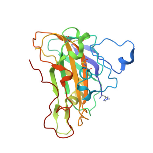
7PU1

High resolution X-ray structure of Thermoascus aurantiacus LPMO


Experimental Data Snapshot

  • Method: X-RAY DIFFRACTION
  • Resolution: 1.06 Å
  • R-Value Free: 0.119 
  • R-Value Work: 0.117 

Starting Model: experimental
View more details

wwPDB Validation   3D Report Full Report


Ligand Structure Quality Assessment 


This is version 1.1 of the entry. See complete history


Literature

Protonation State of an Important Histidine from High Resolution Structures of Lytic Polysaccharide Monooxygenases.

Banerjee, S.Muderspach, S.J.Tandrup, T.Frandsen, K.E.H.Singh, R.K.Ipsen, J.O.Hernandez-Rollan, C.Norholm, M.H.H.Bjerrum, M.J.Johansen, K.S.Lo Leggio, L.

(2022) Biomolecules 12

  • DOI: https://doi.org/10.3390/biom12020194
  • Primary Citation of Related Structures:  
    7PTZ, 7PU1

  • PubMed Abstract: 

    Lytic Polysaccharide Monooxygenases (LPMOs) oxidatively cleave recalcitrant polysaccharides. The mechanism involves (i) reduction of the Cu, (ii) polysaccharide binding, (iii) binding of different oxygen species, and (iv) glycosidic bond cleavage. However, the complete mechanism is poorly understood and may vary across different families and even within the same family. Here, we have investigated the protonation state of a secondary co-ordination sphere histidine, conserved across AA9 family LPMOs that has previously been proposed to be a potential proton donor. Partial unrestrained refinement of newly obtained higher resolution data for two AA9 LPMOs and re-refinement of four additional data sets deposited in the PDB were carried out, where the His was refined without restraints, followed by measurements of the His ring geometrical parameters. This allowed reliable assignment of the protonation state, as also validated by following the same procedure for the His brace, for which the protonation state is predictable. The study shows that this histidine is generally singly protonated at the Nε2 atom, which is close to the oxygen species binding site. Our results indicate robustness of the method. In view of this and other emerging evidence, a role as proton donor during catalysis is unlikely for this His.


  • Organizational Affiliation

    Department of Chemistry, University of Copenhagen, Universitetsparken 5, DK-2100 Copenhagen, Denmark.


Macromolecules
Find similar proteins by:  (by identity cutoff)  |  3D Structure
Entity ID: 1
MoleculeChains Sequence LengthOrganismDetailsImage
Gh61 isozyme aA [auth AAA],
B [auth BBB]
228Thermoascus aurantiacusMutation(s): 0 
EC: 3.2.1.4 (PDB Primary Data), 1.14.99.56 (UniProt)
UniProt
Find proteins for G3XAP7 (Thermoascus aurantiacus)
Explore G3XAP7 
Go to UniProtKB:  G3XAP7
Entity Groups  
Sequence Clusters30% Identity50% Identity70% Identity90% Identity95% Identity100% Identity
UniProt GroupG3XAP7
Glycosylation
Glycosylation Sites: 1
Sequence Annotations
Expand
  • Reference Sequence
Small Molecules
Ligands 3 Unique
IDChains Name / Formula / InChI Key2D Diagram3D Interactions
NAG
Query on NAG

Download Ideal Coordinates CCD File 
D [auth AAA],
N [auth BBB]
2-acetamido-2-deoxy-beta-D-glucopyranose
C8 H15 N O6
OVRNDRQMDRJTHS-FMDGEEDCSA-N
CU
Query on CU

Download Ideal Coordinates CCD File 
C [auth AAA],
O [auth BBB]
COPPER (II) ION
Cu
JPVYNHNXODAKFH-UHFFFAOYSA-N
CL
Query on CL

Download Ideal Coordinates CCD File 
E [auth AAA]
F [auth AAA]
G [auth AAA]
H [auth AAA]
I [auth AAA]
E [auth AAA],
F [auth AAA],
G [auth AAA],
H [auth AAA],
I [auth AAA],
J [auth AAA],
K [auth AAA],
L [auth AAA],
M [auth AAA],
P [auth BBB],
Q [auth BBB],
R [auth BBB],
S [auth BBB],
T [auth BBB],
U [auth BBB],
V [auth BBB],
W [auth BBB],
X [auth BBB],
Y [auth BBB]
CHLORIDE ION
Cl
VEXZGXHMUGYJMC-UHFFFAOYSA-M
Modified Residues  1 Unique
IDChains TypeFormula2D DiagramParent
HIC
Query on HIC
A [auth AAA],
B [auth BBB]
L-PEPTIDE LINKINGC7 H11 N3 O2HIS
Experimental Data & Validation

Experimental Data

  • Method: X-RAY DIFFRACTION
  • Resolution: 1.06 Å
  • R-Value Free: 0.119 
  • R-Value Work: 0.117 
  • Space Group: P 1 21 1
Unit Cell:
Length ( Å )Angle ( ˚ )
a = 37.653α = 90
b = 89.046β = 103.335
c = 70.47γ = 90
Software Package:
Software NamePurpose
REFMACrefinement
XDSdata reduction
TRUNCATEdata scaling
REFMACphasing

Structure Validation

View Full Validation Report


Ligand Structure Quality Assessment 


Entry History & Funding Information

Deposition Data


Funding OrganizationLocationGrant Number
Novo Nordisk FoundationSwedenNNF17SA0027704
Danish Council for Independent ResearchSweden8021-00273B
Swedish Research CouncilSweden2018-04969; 2019-02496

Revision History  (Full details and data files)

  • Version 1.0: 2022-03-16
    Type: Initial release
  • Version 1.1: 2024-01-31
    Changes: Data collection, Derived calculations, Refinement description