7ZTL

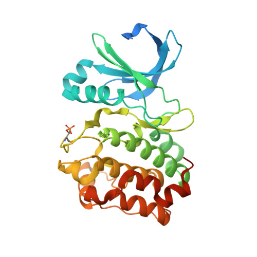
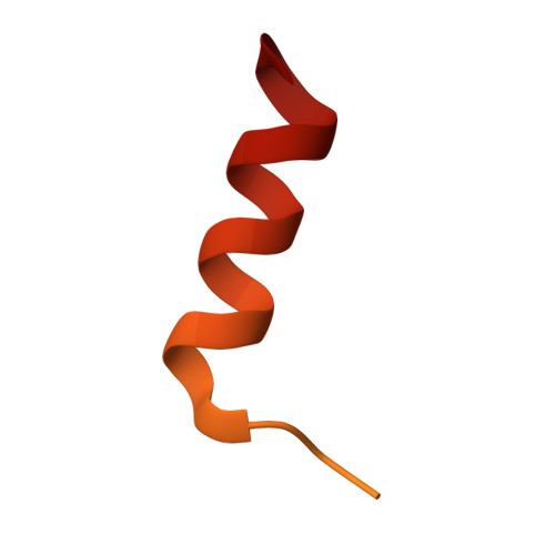
Crystal structure of a covalently linked Aurora-A N-Myc complex


Experimental Data Snapshot

  • Method: X-RAY DIFFRACTION
  • Resolution: 1.90 Å
  • R-Value Free: 0.193 
  • R-Value Work: 0.163 
  • R-Value Observed: 0.165 

Starting Model: experimental
View more details

wwPDB Validation   3D Report Full Report


Ligand Structure Quality Assessment 


This is version 1.3 of the entry. See complete history


Literature

Crystal structure of a covalently linked Aurora-A-MYCN complex.

Diebold, M.Schonemann, L.Eilers, M.Sotriffer, C.Schindelin, H.

(2023) Acta Crystallogr D Struct Biol 79: 1-9

  • DOI: https://doi.org/10.1107/S2059798322011433
  • Primary Citation of Related Structures:  
    7ZTL

  • PubMed Abstract: 

    Formation of the Aurora-A-MYCN complex increases levels of the oncogenic transcription factor MYCN in neuroblastoma cells by abrogating its degradation through the ubiquitin proteasome system. While some small-molecule inhibitors of Aurora-A were shown to destabilize MYCN, clinical trials have not been satisfactory to date. MYCN itself is considered to be `undruggable' due to its large intrinsically disordered regions. Targeting the Aurora-A-MYCN complex rather than Aurora-A or MYCN alone will open new possibilities for drug development and screening campaigns. To overcome the challenges that a ternary system composed of Aurora-A, MYCN and a small molecule entails, a covalently cross-linked construct of the Aurora-A-MYCN complex was designed, expressed and characterized, thus enabling screening and design campaigns to identify selective binders.


  • Organizational Affiliation

    Institute of Pharmacy and Food Chemistry, University of Würzburg, Am Hubland, 97074 Würzburg, Germany.


Macromolecules
Find similar proteins by:  (by identity cutoff)  |  3D Structure
Entity ID: 1
MoleculeChains Sequence LengthOrganismDetailsImage
Aurora kinase A309Homo sapiensMutation(s): 3 
Gene Names: AURKAAIKAIRK1ARK1AURAAYK1BTAKIAK1STK15STK6
EC: 2.7.11.1
UniProt & NIH Common Fund Data Resources
Find proteins for O14965 (Homo sapiens)
Explore O14965 
Go to UniProtKB:  O14965
PHAROS:  O14965
GTEx:  ENSG00000087586 
Entity Groups  
Sequence Clusters30% Identity50% Identity70% Identity90% Identity95% Identity100% Identity
UniProt GroupO14965
Sequence Annotations
Expand
  • Reference Sequence
Find similar proteins by:  (by identity cutoff)  |  3D Structure
Entity ID: 2
MoleculeChains Sequence LengthOrganismDetailsImage
N-myc proto-oncogene protein62Homo sapiensMutation(s): 1 
UniProt & NIH Common Fund Data Resources
Find proteins for P04198 (Homo sapiens)
Explore P04198 
Go to UniProtKB:  P04198
PHAROS:  P04198
GTEx:  ENSG00000134323 
Entity Groups  
Sequence Clusters30% Identity50% Identity70% Identity90% Identity95% Identity100% Identity
UniProt GroupP04198
Sequence Annotations
Expand
  • Reference Sequence
Small Molecules
Ligands 6 Unique
IDChains Name / Formula / InChI Key2D Diagram3D Interactions
ADP
Query on ADP

Download Ideal Coordinates CCD File 
C [auth A]ADENOSINE-5'-DIPHOSPHATE
C10 H15 N5 O10 P2
XTWYTFMLZFPYCI-KQYNXXCUSA-N
JWG (Subject of Investigation/LOI)
Query on JWG

Download Ideal Coordinates CCD File 
K [auth B]4-(3-hydroxy-3-oxopropylamino)-4-oxidanylidene-butanoic acid
C7 H11 N O5
NFQWOKKYYQQDCQ-UHFFFAOYSA-N
BCN
Query on BCN

Download Ideal Coordinates CCD File 
H [auth A]BICINE
C6 H13 N O4
FSVCELGFZIQNCK-UHFFFAOYSA-N
GOL
Query on GOL

Download Ideal Coordinates CCD File 
F [auth A],
G [auth A],
I [auth A]
GLYCEROL
C3 H8 O3
PEDCQBHIVMGVHV-UHFFFAOYSA-N
MG
Query on MG

Download Ideal Coordinates CCD File 
D [auth A],
E [auth A]
MAGNESIUM ION
Mg
JLVVSXFLKOJNIY-UHFFFAOYSA-N
NA
Query on NA

Download Ideal Coordinates CCD File 
J [auth A]SODIUM ION
Na
FKNQFGJONOIPTF-UHFFFAOYSA-N
Modified Residues  1 Unique
IDChains TypeFormula2D DiagramParent
TPO
Query on TPO
A
L-PEPTIDE LINKINGC4 H10 N O6 PTHR
Experimental Data & Validation

Experimental Data

  • Method: X-RAY DIFFRACTION
  • Resolution: 1.90 Å
  • R-Value Free: 0.193 
  • R-Value Work: 0.163 
  • R-Value Observed: 0.165 
  • Space Group: P 32 2 1
Unit Cell:
Length ( Å )Angle ( ˚ )
a = 86.614α = 90
b = 86.614β = 90
c = 91.789γ = 120
Software Package:
Software NamePurpose
PHENIXrefinement
PHASERphasing
Aimlessdata scaling
XDSdata reduction

Structure Validation

View Full Validation Report


Ligand Structure Quality Assessment 


Entry History & Funding Information

Deposition Data


Funding OrganizationLocationGrant Number
German Research Foundation (DFG)GermanyGRK2243

Revision History  (Full details and data files)

  • Version 1.0: 2023-01-18
    Type: Initial release
  • Version 1.1: 2024-01-31
    Changes: Data collection, Refinement description
  • Version 1.2: 2024-04-03
    Changes: Database references
  • Version 1.3: 2024-11-06
    Changes: Structure summary