8FBM

Crystal structure of Cryptosporidium parvum N-myristoyltransferase with bound myristoyl-CoA and inhibitor 1


Experimental Data Snapshot

  • Method: X-RAY DIFFRACTION
  • Resolution: 1.90 Å
  • R-Value Free: 0.190 
  • R-Value Work: 0.148 
  • R-Value Observed: 0.150 

Starting Model: experimental
View more details

wwPDB Validation   3D Report Full Report


Ligand Structure Quality Assessment 


This is version 1.2 of the entry. See complete history


Literature

Identification of and Structural Insights into Hit Compounds Targeting N -Myristoyltransferase for Cryptosporidium Drug Development.

Fenwick, M.K.Reers, A.R.Liu, Y.Zigweid, R.Sankaran, B.Shin, J.Hulverson, M.A.Hammerson, B.Fernandez Alvaro, E.Myler, P.J.Kaushansky, A.Van Voorhis, W.C.Fan, E.Staker, B.L.

(2023) ACS Infect Dis 9: 1821-1833

  • DOI: https://doi.org/10.1021/acsinfecdis.3c00151
  • Primary Citation of Related Structures:  
    8FBM, 8FBT, 8FBU

  • PubMed Abstract: 

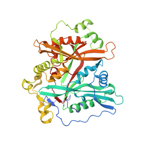
    Each year, approximately 50,000 children under 5 die as a result of diarrhea caused by Cryptosporidium parvum , a protozoan parasite. There are currently no effective drugs or vaccines available to cure or prevent Cryptosporidium infection, and there are limited tools for identifying and validating targets for drug or vaccine development. We previously reported a high throughput screening (HTS) of a large compound library against Plasmodium N -myristoyltransferase (NMT), a validated drug target in multiple protozoan parasite species. To identify molecules that could be effective against Cryptosporidium , we counter-screened hits from the Plasmodium NMT HTS against Cryptosporidium NMT. We identified two potential hit compounds and validated them against Cp NMT to determine if NMT might be an attractive drug target also for Cryptosporidium . We tested the compounds against Cryptosporidium using both cell-based and NMT enzymatic assays. We then determined the crystal structure of Cp NMT bound to Myristoyl-Coenzyme A (MyrCoA) and structures of ternary complexes with MyrCoA and the hit compounds to identify the ligand binding modes. The binding site architectures display different conformational states in the presence of the two inhibitors and provide a basis for rational design of selective inhibitors.


  • Organizational Affiliation

    Seattle Structural Genomics Center for Infectious Disease (SSGCID), Seattle, Washington 98109, United States.


Macromolecules
Find similar proteins by:  (by identity cutoff)  |  3D Structure
Entity ID: 1
MoleculeChains Sequence LengthOrganismDetailsImage
Glycylpeptide N-tetradecanoyltransferase
A, B
431Cryptosporidium parvum Iowa IIMutation(s): 0 
Gene Names: cgd3_320
UniProt
Find proteins for Q5CV46 (Cryptosporidium parvum (strain Iowa II))
Explore Q5CV46 
Go to UniProtKB:  Q5CV46
Entity Groups  
Sequence Clusters30% Identity50% Identity70% Identity90% Identity95% Identity100% Identity
UniProt GroupQ5CV46
Sequence Annotations
Expand
  • Reference Sequence
Small Molecules
Ligands 6 Unique
IDChains Name / Formula / InChI Key2D Diagram3D Interactions
MYA (Subject of Investigation/LOI)
Query on MYA

Download Ideal Coordinates CCD File 
C [auth A],
K [auth B]
TETRADECANOYL-COA
C35 H62 N7 O17 P3 S
DUAFKXOFBZQTQE-QSGBVPJFSA-N
XOF (Subject of Investigation/LOI)
Query on XOF

Download Ideal Coordinates CCD File 
D [auth A],
L [auth B]
2-chloro-5-[ethyl(phenyl)sulfamoyl]-N-[2-(2-oxopyrrolidin-1-yl)phenyl]benzamide
C25 H24 Cl N3 O4 S
DXAJOSDSBRJHSK-UHFFFAOYSA-N
PG4
Query on PG4

Download Ideal Coordinates CCD File 
I [auth A],
M [auth B],
S [auth B]
TETRAETHYLENE GLYCOL
C8 H18 O5
UWHCKJMYHZGTIT-UHFFFAOYSA-N
PEG
Query on PEG

Download Ideal Coordinates CCD File 
E [auth A],
F [auth A],
J [auth A],
R [auth B]
DI(HYDROXYETHYL)ETHER
C4 H10 O3
MTHSVFCYNBDYFN-UHFFFAOYSA-N
GOL
Query on GOL

Download Ideal Coordinates CCD File 
N [auth B]GLYCEROL
C3 H8 O3
PEDCQBHIVMGVHV-UHFFFAOYSA-N
CL
Query on CL

Download Ideal Coordinates CCD File 
G [auth A],
H [auth A],
O [auth B],
P [auth B],
Q [auth B]
CHLORIDE ION
Cl
VEXZGXHMUGYJMC-UHFFFAOYSA-M
Experimental Data & Validation

Experimental Data

  • Method: X-RAY DIFFRACTION
  • Resolution: 1.90 Å
  • R-Value Free: 0.190 
  • R-Value Work: 0.148 
  • R-Value Observed: 0.150 
  • Space Group: P 1 21 1
Unit Cell:
Length ( Å )Angle ( ˚ )
a = 54.036α = 90
b = 88.332β = 97.67
c = 98.322γ = 90
Software Package:
Software NamePurpose
PHENIXrefinement
HKL-2000data reduction
HKL-2000data scaling
MOLREPphasing

Structure Validation

View Full Validation Report


Ligand Structure Quality Assessment 


Entry History & Funding Information

Deposition Data


Funding OrganizationLocationGrant Number
National Institutes of Health/National Institute Of Allergy and Infectious Diseases (NIH/NIAID)United StatesR01AI155536

Revision History  (Full details and data files)

  • Version 1.0: 2023-05-31
    Type: Initial release
  • Version 1.1: 2023-09-20
    Changes: Data collection, Refinement description
  • Version 1.2: 2024-09-04
    Changes: Data collection, Database references, Structure summary