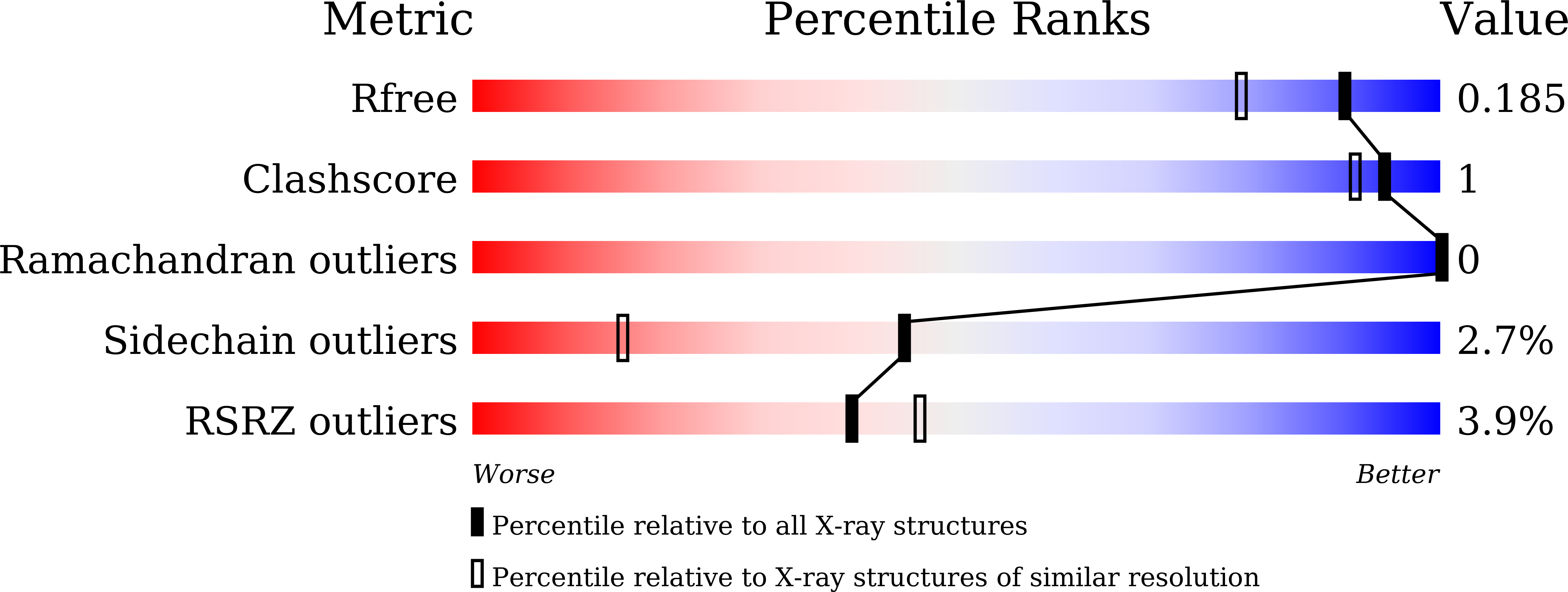
8IW3

crystal structure of SulE mutant


Experimental Data Snapshot

  • Method: X-RAY DIFFRACTION
  • Resolution: 1.56 Å
  • R-Value Free: 0.185 
  • R-Value Work: 0.170 

Starting Model: experimental
View more details

wwPDB Validation   3D Report Full Report


Ligand Structure Quality Assessment 


This is version 1.1 of the entry. See complete history


Literature

Crystal structures of herbicide-detoxifying esterase reveal a lid loop affecting substrate binding and activity.

Liu, B.Wang, W.Qiu, J.Huang, X.Qiu, S.Bao, Y.Xu, S.Ruan, L.Ran, T.He, J.

(2023) Nat Commun 14: 4343-4343

  • DOI: https://doi.org/10.1038/s41467-023-40103-5
  • Primary Citation of Related Structures:  
    7Y0L, 7YD2, 8GOL, 8GOY, 8GP0, 8IVE, 8IVM, 8IVN, 8IVS, 8IVT, 8IW3, 8IW6, 8J7G, 8J7J, 8J7K

  • PubMed Abstract: 

    SulE, an esterase, which detoxifies a variety of sulfonylurea herbicides through de-esterification, provides an attractive approach to remove environmental sulfonylurea herbicides and develop herbicide-tolerant crops. Here, we determined the crystal structures of SulE and an activity improved mutant P44R. Structural analysis revealed that SulE is a dimer with spacious binding pocket accommodating the large sulfonylureas substrate. Particularly, SulE contains a protruding β hairpin with a lid loop covering the active site of the other subunit of the dimer. The lid loop participates in substrate recognition and binding. P44R mutation altered the lid loop flexibility, resulting in the sulfonylurea heterocyclic ring repositioning to a relative stable conformation thus leading to dramatically increased activity. Our work provides important insights into the molecular mechanism of SulE, and establish a solid foundation for further improving the enzyme activity to various sulfonylurea herbicides through rational design.


  • Organizational Affiliation

    Key Laboratory of Agricultural Environmental Microbiology of Ministry of Agriculture, College of Life Sciences, Nanjing Agricultural University, Nanjing, 210095, China.


Macromolecules
Find similar proteins by:  (by identity cutoff)  |  3D Structure
Entity ID: 1
MoleculeChains Sequence LengthOrganismDetailsImage
Alpha/beta fold hydrolase
A, B
370Hansschlegelia zhihuaiaeMutation(s): 2 
Gene Names: EK403_17710
EC: 3.1.1
UniProt
Find proteins for G9I933 (Hansschlegelia zhihuaiae)
Explore G9I933 
Go to UniProtKB:  G9I933
Entity Groups  
Sequence Clusters30% Identity50% Identity70% Identity90% Identity95% Identity100% Identity
UniProt GroupG9I933
Sequence Annotations
Expand
  • Reference Sequence
Small Molecules
Ligands 3 Unique
IDChains Name / Formula / InChI Key2D Diagram3D Interactions
CIE (Subject of Investigation/LOI)
Query on CIE

Download Ideal Coordinates CCD File 
C [auth A],
F [auth B]
2-[[[[(4-CHLORO-6-METHOXY-2-PYRIMIDINYL)AMINO]CARBONYL]AMINO]SULFONYL]BENZOIC ACID ETHYL ESTER
C15 H15 Cl N4 O6 S
NSWAMPCUPHPTTC-UHFFFAOYSA-N
TLA
Query on TLA

Download Ideal Coordinates CCD File 
D [auth A],
G [auth B]
L(+)-TARTARIC ACID
C4 H6 O6
FEWJPZIEWOKRBE-JCYAYHJZSA-N
GOL
Query on GOL

Download Ideal Coordinates CCD File 
E [auth A],
H [auth B]
GLYCEROL
C3 H8 O3
PEDCQBHIVMGVHV-UHFFFAOYSA-N
Experimental Data & Validation

Experimental Data

  • Method: X-RAY DIFFRACTION
  • Resolution: 1.56 Å
  • R-Value Free: 0.185 
  • R-Value Work: 0.170 
  • Space Group: P 1 21 1
Unit Cell:
Length ( Å )Angle ( ˚ )
a = 51.36α = 90
b = 140.03β = 101.41
c = 58.26γ = 90
Software Package:
Software NamePurpose
PHENIXrefinement
XDSdata reduction
SCALAdata scaling
PHASERphasing

Structure Validation

View Full Validation Report


Ligand Structure Quality Assessment 


Entry History & Funding Information

Deposition Data


Funding OrganizationLocationGrant Number
National Natural Science Foundation of China (NSFC)China31970096

Revision History  (Full details and data files)

  • Version 1.0: 2023-08-02
    Type: Initial release
  • Version 1.1: 2024-05-29
    Changes: Data collection