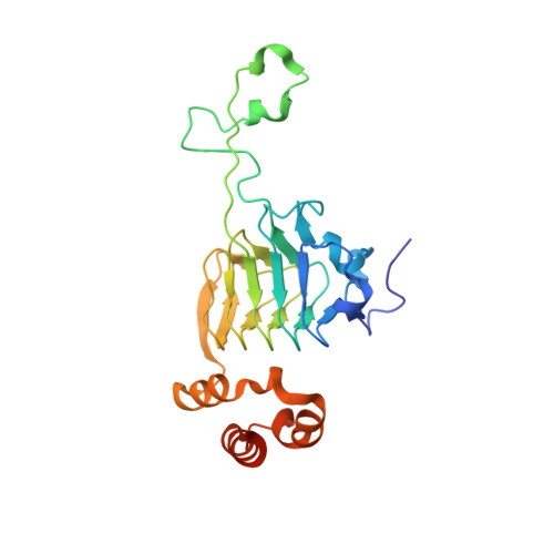
8J40

Crystal Structure of CATB8 in complex with chloramphenicol


Experimental Data Snapshot

  • Method: X-RAY DIFFRACTION
  • Resolution: 1.57 Å
  • R-Value Free: 0.163 
  • R-Value Work: 0.156 
  • R-Value Observed: 0.156 

Starting Model: experimental
View more details

wwPDB Validation   3D Report Full Report


Ligand Structure Quality Assessment 


This is version 1.1 of the entry. See complete history


Literature

Chloramphenicol Binding Sites of Acinetobacter baumannii Chloramphenicol Acetyltransferase CatB8.

Liao, J.Qi, Q.Kuang, L.Zhou, Y.Xiao, Q.Liu, T.Wang, X.Guo, L.Jiang, Y.

(2024) ACS Infect Dis 10: 870-878

  • DOI: https://doi.org/10.1021/acsinfecdis.3c00359
  • Primary Citation of Related Structures:  
    8J40

  • PubMed Abstract: 

    Acinetobacter baumannii is a multidrug-resistant pathogen that has become one of the most challenging pathogens in global healthcare. Several antibiotic-resistant genes, including catB8 , have been identified in the A. baumannii genome. CatB8 protein, one of the chloramphenicol acetyltransferases (Cats), is encoded by the catB8 gene. Cats can convert chloramphenicol (chl) to 3-acetyl-chl, leading to bacterial resistance to chl. Here, we present the high-resolution cocrystal structure of CatB8 with chl. The structure that we resolved showed that each monomer of CatB8 binds to four chl molecules, while its homologous protein only binds to one chl molecule. One of the newly discovered chl binding site overlaps with the site of another substrate, acetyl-CoA. Through structure-based biochemical analyses, we identified key residues for chl recruiting and acetylation of chl in CatB8. Our work is of significant importance for understanding the drug resistance of A. baumannii and the effectiveness of antibiotic treatment.


  • Organizational Affiliation

    Department of Laboratory Medicine, West China Second University Hospital, Sichuan University, Chengdu 610041, Sichuan, China.


Macromolecules
Find similar proteins by:  (by identity cutoff)  |  3D Structure
Entity ID: 1
MoleculeChains Sequence LengthOrganismDetailsImage
CatB8218Acinetobacter baumanniiMutation(s): 0 
Gene Names: catB8
EC: 2.3.1.28
UniProt
Find proteins for Q6Q627 (Acinetobacter baumannii)
Explore Q6Q627 
Go to UniProtKB:  Q6Q627
Entity Groups  
Sequence Clusters30% Identity50% Identity70% Identity90% Identity95% Identity100% Identity
UniProt GroupQ6Q627
Sequence Annotations
Expand
  • Reference Sequence
Small Molecules
Ligands 3 Unique
IDChains Name / Formula / InChI Key2D Diagram3D Interactions
CLM (Subject of Investigation/LOI)
Query on CLM

Download Ideal Coordinates CCD File 
B [auth A],
C [auth A],
D [auth A],
E [auth A]
CHLORAMPHENICOL
C11 H12 Cl2 N2 O5
WIIZWVCIJKGZOK-RKDXNWHRSA-N
SO4
Query on SO4

Download Ideal Coordinates CCD File 
J [auth A]SULFATE ION
O4 S
QAOWNCQODCNURD-UHFFFAOYSA-L
GOL
Query on GOL

Download Ideal Coordinates CCD File 
F [auth A],
G [auth A],
H [auth A],
I [auth A]
GLYCEROL
C3 H8 O3
PEDCQBHIVMGVHV-UHFFFAOYSA-N
Experimental Data & Validation

Experimental Data

  • Method: X-RAY DIFFRACTION
  • Resolution: 1.57 Å
  • R-Value Free: 0.163 
  • R-Value Work: 0.156 
  • R-Value Observed: 0.156 
  • Space Group: I 2 3
Unit Cell:
Length ( Å )Angle ( ˚ )
a = 128.548α = 90
b = 128.548β = 90
c = 128.548γ = 90
Software Package:
Software NamePurpose
PHENIXrefinement
HKL-2000data reduction
HKL-2000data scaling
PHASERphasing

Structure Validation

View Full Validation Report


Ligand Structure Quality Assessment 


Entry History & Funding Information

Deposition Data


Funding OrganizationLocationGrant Number
National Natural Science Foundation of China (NSFC)China2019YJ0083

Revision History  (Full details and data files)

  • Version 1.0: 2024-02-28
    Type: Initial release
  • Version 1.1: 2024-03-20
    Changes: Database references