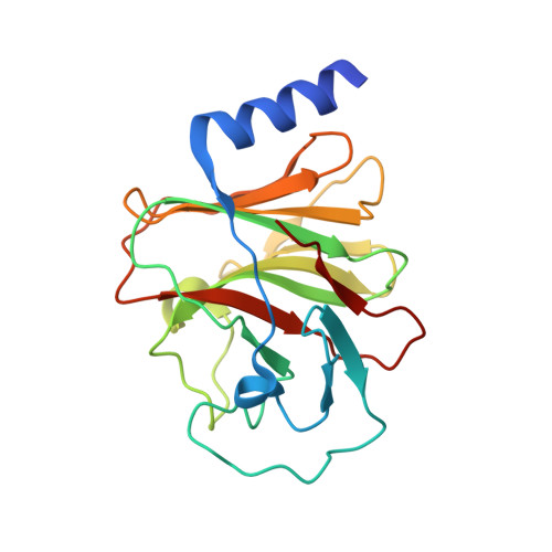
Phosphoantigens glue butyrophilin 3A1 and 2A1 to activate V gamma 9V delta 2 T cells.
Yuan, L., Ma, X., Yang, Y., Qu, Y., Li, X., Zhu, X., Ma, W., Duan, J., Xue, J., Yang, H., Huang, J.W., Yi, S., Zhang, M., Cai, N., Zhang, L., Ding, Q., Lai, K., Liu, C., Zhang, L., Liu, X., Yao, Y., Zhou, S., Li, X., Shen, P., Chang, Q., Malwal, S.R., He, Y., Li, W., Chen, C., Chen, C.C., Oldfield, E., Guo, R.T., Zhang, Y.(2023) Nature 621: 840-848
- PubMed: 37674084
- DOI: https://doi.org/10.1038/s41586-023-06525-3
- Primary Citation of Related Structures:
8HJT, 8IGT, 8IH4, 8IXV, 8IZE, 8IZG, 8JY9, 8JYA, 8JYB, 8JYC, 8JYE, 8JYF - PubMed Abstract:
In both cancer and infections, diseased cells are presented to human Vγ9Vδ2 T cells through an 'inside out' signalling process whereby structurally diverse phosphoantigen (pAg) molecules are sensed by the intracellular domain of butyrophilin BTN3A1 1-4 . Here we show how-in both humans and alpaca-multiple pAgs function as 'molecular glues' to promote heteromeric association between the intracellular domains of BTN3A1 and the structurally similar butyrophilin BTN2A1. X-ray crystallography studies visualized that engagement of BTN3A1 with pAgs forms a composite interface for direct binding to BTN2A1, with various pAg molecules each positioned at the centre of the interface and gluing the butyrophilins with distinct affinities. Our structural insights guided mutagenesis experiments that led to disruption of the intracellular BTN3A1-BTN2A1 association, abolishing pAg-mediated Vγ9Vδ2 T cell activation. Analyses using structure-based molecular-dynamics simulations, 19 F-NMR investigations, chimeric receptor engineering and direct measurement of intercellular binding force revealed how pAg-mediated BTN2A1 association drives BTN3A1 intracellular fluctuations outwards in a thermodynamically favourable manner, thereby enabling BTN3A1 to push off from the BTN2A1 ectodomain to initiate T cell receptor-mediated γδ T cell activation. Practically, we harnessed the molecular-glue model for immunotherapeutics design, demonstrating chemical principles for developing both small-molecule activators and inhibitors of human γδ T cell function.
Organizational Affiliation:
Tsinghua-Peking Center for Life Sciences, State Key Laboratory of Membrane Biology, School of Pharmaceutical Sciences, Tsinghua University, Beijing, China.


