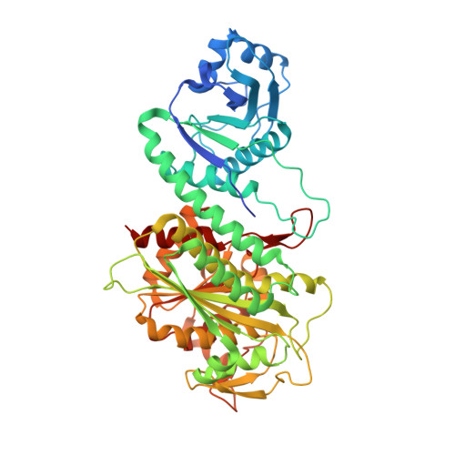
8PZM

Intracellular leucine aminopeptidase of Pseudomonas aeruginosa PA14 bound to bestatin inhibitor and manganese


Experimental Data Snapshot

  • Method: X-RAY DIFFRACTION
  • Resolution: 1.70 Å
  • R-Value Free: 0.189 
  • R-Value Work: 0.170 
  • R-Value Observed: 0.171 

Starting Model: experimental
View more details

wwPDB Validation   3D Report Full Report


Ligand Structure Quality Assessment 


This is version 1.1 of the entry. See complete history


Literature

Unveiling the Catalytic Mechanism of a Processive Metalloaminopeptidase.

Simpson, M.C.Harding, C.J.Czekster, R.M.Remmel, L.Bode, B.E.Czekster, C.M.

(2023) Biochemistry 62: 3188-3205

  • DOI: https://doi.org/10.1021/acs.biochem.3c00420
  • Primary Citation of Related Structures:  
    8PZ0, 8PZM, 8PZY

  • PubMed Abstract: 

    Intracellular leucine aminopeptidases (PepA) are metalloproteases from the family M17. These enzymes catalyze peptide bond cleavage, removing N-terminal residues from peptide and protein substrates, with consequences for protein homeostasis and quality control. While general mechanistic studies using model substrates have been conducted on PepA enzymes from various organisms, specific information about their substrate preferences and promiscuity, choice of metal, activation mechanisms, and the steps that limit steady-state turnover remain unexplored. Here, we dissected the catalytic and chemical mechanisms of Pa PepA: a leucine aminopeptidase from Pseudomonas aeruginosa . Cleavage assays using peptides and small-molecule substrate mimics allowed us to propose a mechanism for catalysis. Steady-state and pre-steady-state kinetics, pH rate profiles, solvent kinetic isotope effects, and biophysical techniques were used to evaluate metal binding and activation. This revealed that metal binding to a tight affinity site is insufficient for enzyme activity; binding to a weaker affinity site is essential for catalysis. Progress curves for peptide hydrolysis and crystal structures of free and inhibitor-bound Pa PepA revealed that Pa PepA cleaves peptide substrates in a processive manner. We propose three distinct modes for activity regulation: tight packing of Pa PepA in a hexameric assembly controls substrate length and reaction processivity; the product leucine acts as an inhibitor, and the high concentration of metal ions required for activation limits catalytic turnover. Our work uncovers catalysis by a metalloaminopeptidase, revealing the intricacies of metal activation and substrate selection. This will pave the way for a deeper understanding of metalloenzymes and processive peptidases/proteases.


  • Organizational Affiliation

    School of Biology, University of St Andrews, North Haugh, Biomolecular Sciences Building, KY16 9ST, Saint Andrews, United Kingdom.


Macromolecules
Find similar proteins by:  (by identity cutoff)  |  3D Structure
Entity ID: 1
MoleculeChains Sequence LengthOrganismDetailsImage
Probable cytosol aminopeptidase517Pseudomonas aeruginosa PA14Mutation(s): 0 
Gene Names: pepA
EC: 3.4.11.1 (UniProt), 3.4.11.10 (UniProt)
UniProt
Find proteins for Q02RY8 (Pseudomonas aeruginosa (strain UCBPP-PA14))
Explore Q02RY8 
Go to UniProtKB:  Q02RY8
Entity Groups  
Sequence Clusters30% Identity50% Identity70% Identity90% Identity95% Identity100% Identity
UniProt GroupQ02RY8
Sequence Annotations
Expand
  • Reference Sequence
Small Molecules
Ligands 4 Unique
IDChains Name / Formula / InChI Key2D Diagram3D Interactions
BES (Subject of Investigation/LOI)
Query on BES

Download Ideal Coordinates CCD File 
B [auth A]2-(3-AMINO-2-HYDROXY-4-PHENYL-BUTYRYLAMINO)-4-METHYL-PENTANOIC ACID
C16 H24 N2 O4
VGGGPCQERPFHOB-RDBSUJKOSA-N
EDO
Query on EDO

Download Ideal Coordinates CCD File 
C [auth A],
E [auth A]
1,2-ETHANEDIOL
C2 H6 O2
LYCAIKOWRPUZTN-UHFFFAOYSA-N
BCT (Subject of Investigation/LOI)
Query on BCT

Download Ideal Coordinates CCD File 
D [auth A]BICARBONATE ION
C H O3
BVKZGUZCCUSVTD-UHFFFAOYSA-M
MN
Query on MN

Download Ideal Coordinates CCD File 
F [auth A],
G [auth A]
MANGANESE (II) ION
Mn
WAEMQWOKJMHJLA-UHFFFAOYSA-N
Experimental Data & Validation

Experimental Data

  • Method: X-RAY DIFFRACTION
  • Resolution: 1.70 Å
  • R-Value Free: 0.189 
  • R-Value Work: 0.170 
  • R-Value Observed: 0.171 
  • Space Group: P 63 2 2
Unit Cell:
Length ( Å )Angle ( ˚ )
a = 182.05α = 90
b = 182.05β = 90
c = 87.57γ = 120
Software Package:
Software NamePurpose
PHENIXrefinement
xia2data scaling
xia2data reduction
PHASERphasing

Structure Validation

View Full Validation Report


Ligand Structure Quality Assessment 


Entry History & Funding Information

Deposition Data


Funding OrganizationLocationGrant Number
Wellcome TrustUnited Kingdom210486/Z/18/Z

Revision History  (Full details and data files)

  • Version 1.0: 2023-11-15
    Type: Initial release
  • Version 1.1: 2023-12-06
    Changes: Database references