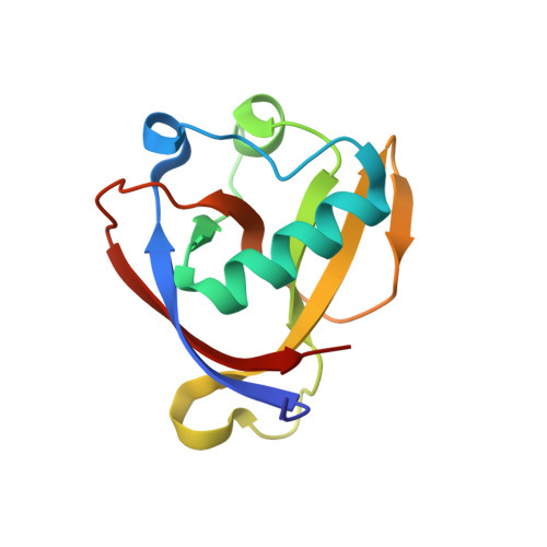
High-confidence placement of low-occupancy fragments into electron density using the anomalous signal of sulfur and halogen atoms.
Ma, S., Damfo, S., Bowler, M.W., Mykhaylyk, V., Kozielski, F.(2024) Acta Crystallogr D Struct Biol 80: 451-463
- PubMed: 38841886
- DOI: https://doi.org/10.1107/S2059798324004480
- Primary Citation of Related Structures:
8RF2, 8RF3, 8RF4, 8RF5, 8RF6, 8RF8, 8RFC, 8RFD, 8RFF - PubMed Abstract:
Fragment-based drug design using X-ray crystallography is a powerful technique to enable the development of new lead compounds, or probe molecules, against biological targets. This study addresses the need to determine fragment binding orientations for low-occupancy fragments with incomplete electron density, an essential step before further development of the molecule. Halogen atoms play multiple roles in drug discovery due to their unique combination of electronegativity, steric effects and hydrophobic properties. Fragments incorporating halogen atoms serve as promising starting points in hit-to-lead development as they often establish halogen bonds with target proteins, potentially enhancing binding affinity and selectivity, as well as counteracting drug resistance. Here, the aim was to unambiguously identify the binding orientations of fragment hits for SARS-CoV-2 nonstructural protein 1 (nsp1) which contain a combination of sulfur and/or chlorine, bromine and iodine substituents. The binding orientations of carefully selected nsp1 analogue hits were focused on by employing their anomalous scattering combined with Pan-Dataset Density Analysis (PanDDA). Anomalous difference Fourier maps derived from the diffraction data collected at both standard and long-wavelength X-rays were compared. The discrepancies observed in the maps of iodine-containing fragments collected at different energies were attributed to site-specific radiation-damage stemming from the strong X-ray absorption of I atoms, which is likely to cause cleavage of the C-I bond. A reliable and effective data-collection strategy to unambiguously determine the binding orientations of low-occupancy fragments containing sulfur and/or halogen atoms while mitigating radiation damage is presented.
Organizational Affiliation:
School of Pharmacy, University College London, 29-39 Brunswick Square, London WC1N 1AX, United Kingdom.


