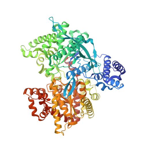
8RVK

Maltodextrin phosphorylase (MalP) in complex with a alpha-1,2-cyclophellitol analogue


Experimental Data Snapshot

  • Method: X-RAY DIFFRACTION
  • Resolution: 2.18 Å
  • R-Value Free: 0.229 
  • R-Value Work: 0.177 

Starting Model: experimental
View more details

wwPDB Validation   3D Report Full Report


Ligand Structure Quality Assessment 


This is version 1.1 of the entry. See complete history


Literature

Conformational and Electronic Variations in 1,2- and 1,5a-Cyclophellitols and their Impact on Retaining alpha-Glucosidase Inhibition.

Ofman, T.P.Heming, J.J.A.Nin-Hill, A.Kullmer, F.Moran, E.Bennett, M.Steneker, R.Klein, A.M.Ruijgrok, G.Kok, K.Armstrong, Z.W.B.Aerts, J.M.F.G.van der Marel, G.A.Rovira, C.Davies, G.J.Artola, M.Codee, J.D.C.Overkleeft, H.S.

(2024) Chemistry 30: e202400723-e202400723

  • DOI: https://doi.org/10.1002/chem.202400723
  • Primary Citation of Related Structures:  
    8RVK, 8RW3

  • PubMed Abstract: 

    Glycoside hydrolases (glycosidases) take part in myriad biological processes and are important therapeutic targets. Competitive and mechanism-based inhibitors are useful tools to dissect their biological role and comprise a good starting point for drug discovery. The natural product, cyclophellitol, a mechanism-based, covalent and irreversible retaining β-glucosidase inhibitor has inspired the design of diverse α- and β-glycosidase inhibitor and activity-based probe scaffolds. Here, we sought to deepen our understanding of the structural and functional requirements of cyclophellitol-type compounds for effective human α-glucosidase inhibition. We synthesized a comprehensive set of α-configured 1,2- and 1,5a-cyclophellitol analogues bearing a variety of electrophilic traps. The inhibitory potency of these compounds was assessed towards both lysosomal and ER retaining α-glucosidases. These studies revealed the 1,5a-cyclophellitols to be the most potent retaining α-glucosidase inhibitors, with the nature of the electrophile determining inhibitory mode of action (covalent or non-covalent). DFT calculations support the ability of the 1,5a-cyclophellitols, but not the 1,2-congeners, to adopt conformations that mimic either the Michaelis complex or transition state of α-glucosidases.


  • Organizational Affiliation

    Leiden Institute of Chemistry, Leiden University, Einsteinweg 55, 2333 CC, Leiden, The Netherlands.


Macromolecules
Find similar proteins by:  (by identity cutoff)  |  3D Structure
Entity ID: 1
MoleculeChains Sequence LengthOrganismDetailsImage
Maltodextrin phosphorylase
A, B
797Escherichia coli K-12Mutation(s): 0 
Gene Names: malPb3417JW5689
EC: 2.4.1.1
UniProt
Find proteins for P00490 (Escherichia coli (strain K12))
Explore P00490 
Go to UniProtKB:  P00490
Entity Groups  
Sequence Clusters30% Identity50% Identity70% Identity90% Identity95% Identity100% Identity
UniProt GroupP00490
Sequence Annotations
Expand
  • Reference Sequence
Small Molecules
Ligands 4 Unique
IDChains Name / Formula / InChI Key2D Diagram3D Interactions
A1H3V (Subject of Investigation/LOI)
Query on A1H3V

Download Ideal Coordinates CCD File 
C [auth A](3~{a}~{R},4~{R},5~{R},6~{R},7~{a}~{S})-6-(hydroxymethyl)-4,5-bis(oxidanyl)-3~{a},4,5,6,7,7~{a}-hexahydro-3~{H}-1,3-benzoxazol-2-one
C8 H13 N O5
MSEYXKZOSYTDQB-VOQCIKJUSA-N
PEG
Query on PEG

Download Ideal Coordinates CCD File 
CA [auth B]
F [auth A]
H [auth A]
T [auth A]
UA [auth B]
CA [auth B],
F [auth A],
H [auth A],
T [auth A],
UA [auth B],
VA [auth B],
WA [auth B]
DI(HYDROXYETHYL)ETHER
C4 H10 O3
MTHSVFCYNBDYFN-UHFFFAOYSA-N
SO4
Query on SO4

Download Ideal Coordinates CCD File 
AA [auth A]
AB [auth B]
BA [auth A]
BB [auth B]
CB [auth B]
AA [auth A],
AB [auth B],
BA [auth A],
BB [auth B],
CB [auth B],
DB [auth B],
EB [auth B],
FB [auth B],
GB [auth B],
HB [auth B],
U [auth A],
V [auth A],
W [auth A],
X [auth A],
XA [auth B],
Y [auth A],
YA [auth B],
Z [auth A],
ZA [auth B]
SULFATE ION
O4 S
QAOWNCQODCNURD-UHFFFAOYSA-L
EDO
Query on EDO

Download Ideal Coordinates CCD File 
D [auth A]
DA [auth B]
E [auth A]
EA [auth B]
FA [auth B]
D [auth A],
DA [auth B],
E [auth A],
EA [auth B],
FA [auth B],
G [auth A],
GA [auth B],
HA [auth B],
I [auth A],
IA [auth B],
J [auth A],
JA [auth B],
K [auth A],
KA [auth B],
L [auth A],
LA [auth B],
M [auth A],
MA [auth B],
N [auth A],
NA [auth B],
O [auth A],
OA [auth B],
P [auth A],
PA [auth B],
Q [auth A],
QA [auth B],
R [auth A],
RA [auth B],
S [auth A],
SA [auth B],
TA [auth B]
1,2-ETHANEDIOL
C2 H6 O2
LYCAIKOWRPUZTN-UHFFFAOYSA-N
Modified Residues  1 Unique
IDChains TypeFormula2D DiagramParent
LLP
Query on LLP
A, B
L-PEPTIDE LINKINGC14 H22 N3 O7 PLYS
Experimental Data & Validation

Experimental Data

  • Method: X-RAY DIFFRACTION
  • Resolution: 2.18 Å
  • R-Value Free: 0.229 
  • R-Value Work: 0.177 
  • Space Group: P 21 21 21
Unit Cell:
Length ( Å )Angle ( ˚ )
a = 110.498α = 90
b = 119.79β = 90
c = 150.75γ = 90
Software Package:
Software NamePurpose
REFMACrefinement
DIALSdata scaling
Cootmodel building
MOLREPphasing
xia2data reduction

Structure Validation

View Full Validation Report


Ligand Structure Quality Assessment 


Entry History & Funding Information

Deposition Data


Funding OrganizationLocationGrant Number
European Research Council (ERC)European Union951231

Revision History  (Full details and data files)

  • Version 1.0: 2024-05-15
    Type: Initial release
  • Version 1.1: 2024-06-12
    Changes: Database references