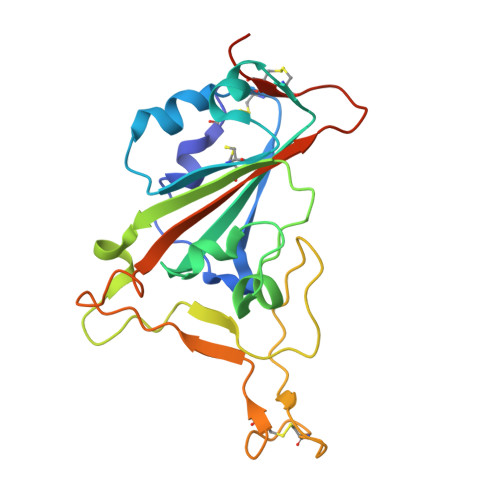
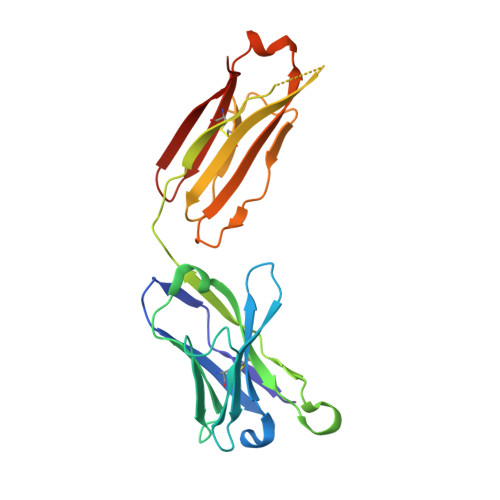
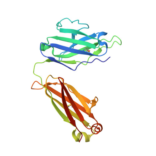
8SDH

Crystal structure of SARS-CoV-2 receptor binding domain in complex with neutralizing antibody CC25.56


Experimental Data Snapshot

  • Method: X-RAY DIFFRACTION
  • Resolution: 2.84 Å
  • R-Value Free: 0.283 
  • R-Value Work: 0.246 
  • R-Value Observed: 0.248 

Starting Model: experimental
View more details

wwPDB Validation   3D Report Full Report


Ligand Structure Quality Assessment 


This is version 1.2 of the entry. See complete history


Literature

Broadly neutralizing antibodies targeting a conserved silent face of spike RBD resist extreme SARS-CoV-2 antigenic drift.

Song, G.Yuan, M.Liu, H.Capozzola, T.Lin, R.N.Torres, J.L.He, W.T.Musharrafieh, R.Dueker, K.Zhou, P.Callaghan, S.Mishra, N.Yong, P.Anzanello, F.Avillion, G.Vo, A.L.Li, X.Makhdoomi, M.Feng, Z.Zhu, X.Peng, L.Nemazee, D.Safonova, Y.Briney, B.Ward, A.B.Burton, D.R.Wilson, I.A.Andrabi, R.

(2023) bioRxiv 

  • DOI: https://doi.org/10.1101/2023.04.26.538488
  • Primary Citation of Related Structures:  
    8SDF, 8SDG, 8SDH

  • PubMed Abstract: 

    Developing broad coronavirus vaccines requires identifying and understanding the molecular basis of broadly neutralizing antibody (bnAb) spike sites. In our previous work, we identified sarbecovirus spike RBD group 1 and 2 bnAbs. We have now shown that many of these bnAbs can still neutralize highly mutated SARS-CoV-2 variants, including the XBB.1.5. Structural studies revealed that group 1 bnAbs use recurrent germline-encoded CDRH3 features to interact with a conserved RBD region that overlaps with class 4 bnAb site. Group 2 bnAbs recognize a less well-characterized "site V" on the RBD and destabilize spike trimer. The site V has remained largely unchanged in SARS-CoV-2 variants and is highly conserved across diverse sarbecoviruses, making it a promising target for broad coronavirus vaccine development. Our findings suggest that targeted vaccine strategies may be needed to induce effective B cell responses to escape resistant subdominant spike RBD bnAb sites.


  • Organizational Affiliation

    Department of Immunology and Microbiology, The Scripps Research Institute, La Jolla, CA 92037, USA.


Macromolecules
Find similar proteins by:  (by identity cutoff)  |  3D Structure
Entity ID: 1
MoleculeChains Sequence LengthOrganismDetailsImage
Spike protein S1
A, B
205Severe acute respiratory syndrome coronavirus 2Mutation(s): 0 
UniProt
Find proteins for P0DTC2 (Severe acute respiratory syndrome coronavirus 2)
Explore P0DTC2 
Go to UniProtKB:  P0DTC2
Entity Groups  
Sequence Clusters30% Identity50% Identity70% Identity90% Identity95% Identity100% Identity
UniProt GroupP0DTC2
Glycosylation
Glycosylation Sites: 1Go to GlyGen: P0DTC2-1
Sequence Annotations
Expand
  • Reference Sequence
Find similar proteins by:  (by identity cutoff)  |  3D Structure
Entity ID: 2
MoleculeChains Sequence LengthOrganismDetailsImage
Neutralizing antibody CC25.56 Heavy ChainC [auth H],
E [auth C]
223Homo sapiensMutation(s): 0 
Entity Groups  
Sequence Clusters30% Identity50% Identity70% Identity90% Identity95% Identity100% Identity
Sequence Annotations
Expand
  • Reference Sequence
Find similar proteins by:  (by identity cutoff)  |  3D Structure
Entity ID: 3
MoleculeChains Sequence LengthOrganismDetailsImage
Neutralizing antibody CC25.56 Light ChainD [auth L],
F [auth D]
214Homo sapiensMutation(s): 0 
Entity Groups  
Sequence Clusters30% Identity50% Identity70% Identity90% Identity95% Identity100% Identity
Sequence Annotations
Expand
  • Reference Sequence
Oligosaccharides

Help

Entity ID: 4
MoleculeChains Length2D Diagram Glycosylation3D Interactions
2-acetamido-2-deoxy-beta-D-glucopyranose-(1-4)-2-acetamido-2-deoxy-beta-D-glucopyranoseG [auth E]2N-Glycosylation
Glycosylation Resources
GlyTouCan:  G42666HT
GlyCosmos:  G42666HT
GlyGen:  G42666HT
Small Molecules
Ligands 1 Unique
IDChains Name / Formula / InChI Key2D Diagram3D Interactions
NAG
Query on NAG

Download Ideal Coordinates CCD File 
H [auth B]2-acetamido-2-deoxy-beta-D-glucopyranose
C8 H15 N O6
OVRNDRQMDRJTHS-FMDGEEDCSA-N
Experimental Data & Validation

Experimental Data

  • Method: X-RAY DIFFRACTION
  • Resolution: 2.84 Å
  • R-Value Free: 0.283 
  • R-Value Work: 0.246 
  • R-Value Observed: 0.248 
  • Space Group: C 1 2 1
Unit Cell:
Length ( Å )Angle ( ˚ )
a = 171.052α = 90
b = 105.586β = 117.31
c = 98.993γ = 90
Software Package:
Software NamePurpose
PHENIXrefinement
XDSdata reduction
XDSdata scaling
PHASERphasing

Structure Validation

View Full Validation Report


Ligand Structure Quality Assessment 


Entry History & Funding Information

Deposition Data


Funding OrganizationLocationGrant Number
National Institutes of Health/National Institute Of Allergy and Infectious Diseases (NIH/NIAID)United StatesR01AI170928
National Institutes of Health/National Institute Of Allergy and Infectious Diseases (NIH/NIAID)United StatesCHAVD UM1 AI44462
Bill & Melinda Gates FoundationUnited StatesINV-004923

Revision History  (Full details and data files)

  • Version 1.0: 2024-08-07
    Type: Initial release
  • Version 1.1: 2024-09-11
    Changes: Derived calculations
  • Version 1.2: 2024-11-13
    Changes: Structure summary