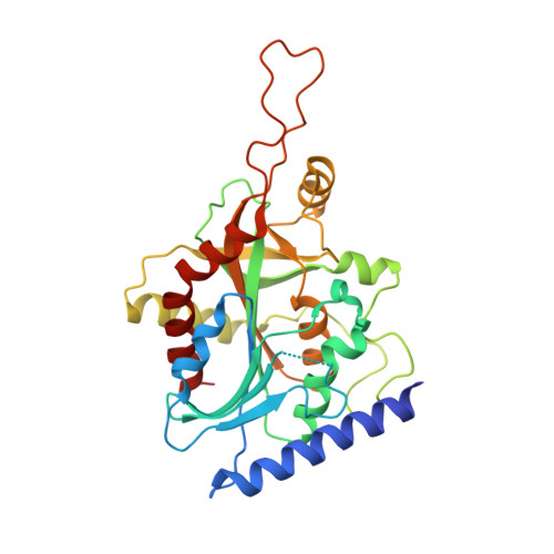
8SWR

Structure of K. lactis PNP S42E variant bound to transition state analog DADMe-IMMUCILLIN G and sulfate


Experimental Data Snapshot

  • Method: X-RAY DIFFRACTION
  • Resolution: 2.30 Å
  • R-Value Free: 0.236 
  • R-Value Work: 0.201 
  • R-Value Observed: 0.203 

Starting Model: experimental
View more details

wwPDB Validation   3D Report Full Report


Ligand Structure Quality Assessment 


This is version 1.2 of the entry. See complete history


Literature

Phosphate Binding in PNP Alters Transition-State Analogue Affinity and Subunit Cooperativity.

Minnow, Y.V.T.Schramm, V.L.Almo, S.C.Ghosh, A.

(2023) Biochemistry 62: 3116-3125

  • DOI: https://doi.org/10.1021/acs.biochem.3c00264
  • Primary Citation of Related Structures:  
    8SWP, 8SWQ, 8SWR, 8SWS, 8SWT, 8SWU

  • PubMed Abstract: 

    Purine nucleoside phosphorylases (PNPs) catalyze the phosphorolysis of 6-oxypurine nucleosides with an HPO 4 2- dianion nucleophile. Nucleosides and phosphate occupy distinct pockets in the PNP active site. Evaluation of the HPO 4 2- site by mutagenesis, cooperative binding studies, and thermodynamic and structural analysis demonstrate that alterations in the HPO 4 2- binding site can render PNP inactive and significantly impact subunit cooperativity and binding to transition-state analogue inhibitors. Cooperative interactions between the cationic transition-state analogue and the anionic HPO 4 2- nucleophile demonstrate the importance of reforming the transition-state ensemble for optimal inhibition with transition-state analogues. Altered phosphate binding in the catalytic site mutants helps to explain one of the known lethal PNP deficiency syndromes in humans.


  • Organizational Affiliation

    Department of Biochemistry, Albert Einstein College of Medicine, 1300 Morris Park Avenue, Bronx, New York 10461, United States.


Macromolecules
Find similar proteins by:  (by identity cutoff)  |  3D Structure
Entity ID: 1
MoleculeChains Sequence LengthOrganismDetailsImage
Purine nucleoside phosphorylase
A, B, C, D, E
A, B, C, D, E, F
308Kluyveromyces lactis NRRL Y-1140Mutation(s): 1 
Gene Names: KLLA0_C16621g
EC: 2.4.2.1
UniProt
Find proteins for Q6CSZ6 (Kluyveromyces lactis (strain ATCC 8585 / CBS 2359 / DSM 70799 / NBRC 1267 / NRRL Y-1140 / WM37))
Explore Q6CSZ6 
Go to UniProtKB:  Q6CSZ6
Entity Groups  
Sequence Clusters30% Identity50% Identity70% Identity90% Identity95% Identity100% Identity
UniProt GroupQ6CSZ6
Sequence Annotations
Expand
  • Reference Sequence
Small Molecules
Ligands 3 Unique
IDChains Name / Formula / InChI Key2D Diagram3D Interactions
IM5 (Subject of Investigation/LOI)
Query on IM5

Download Ideal Coordinates CCD File 
G [auth A],
I [auth B],
J [auth D]
2-amino-7-{[(3R,4R)-3-hydroxy-4-(hydroxymethyl)pyrrolidin-1-yl]methyl}-3,5-dihydro-4H-pyrrolo[3,2-d]pyrimidin-4-one
C12 H17 N5 O3
GSPTUGDLYPMLCQ-SFYZADRCSA-N
GUN
Query on GUN

Download Ideal Coordinates CCD File 
L [auth E]GUANINE
C5 H5 N5 O
UYTPUPDQBNUYGX-UHFFFAOYSA-N
SO4 (Subject of Investigation/LOI)
Query on SO4

Download Ideal Coordinates CCD File 
H [auth A],
K [auth D]
SULFATE ION
O4 S
QAOWNCQODCNURD-UHFFFAOYSA-L
Experimental Data & Validation

Experimental Data

  • Method: X-RAY DIFFRACTION
  • Resolution: 2.30 Å
  • R-Value Free: 0.236 
  • R-Value Work: 0.201 
  • R-Value Observed: 0.203 
  • Space Group: P 1 21 1
Unit Cell:
Length ( Å )Angle ( ˚ )
a = 89.43α = 90
b = 111.005β = 108.3
c = 97.87γ = 90
Software Package:
Software NamePurpose
PHENIXrefinement
HKL-3000data scaling
CBASSdata collection
MOSFLMdata reduction
PHASERphasing

Structure Validation

View Full Validation Report


Ligand Structure Quality Assessment 


Entry History & Funding Information

Deposition Data


Funding OrganizationLocationGrant Number
National Institutes of Health/National Institute of General Medical Sciences (NIH/NIGMS)United StatesGM041916
National Institutes of Health/National Cancer Institute (NIH/NCI)United StatesCA013330

Revision History  (Full details and data files)

  • Version 1.0: 2023-10-18
    Type: Initial release
  • Version 1.1: 2023-10-25
    Changes: Database references
  • Version 1.2: 2023-11-22
    Changes: Database references