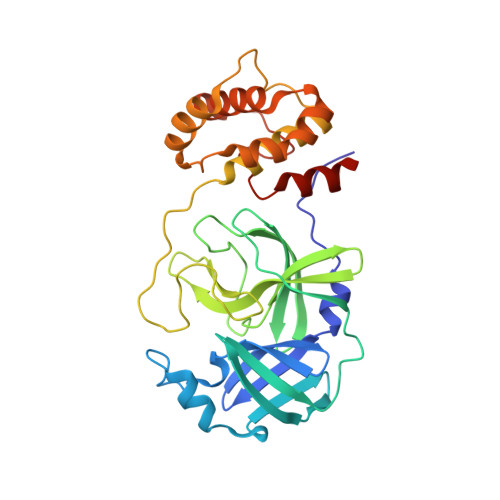
8UHO

Crystal structure of SARS CoV-2 3CL protease in complex with GSK4365096A


Experimental Data Snapshot

  • Method: X-RAY DIFFRACTION
  • Resolution: 2.02 Å
  • R-Value Free: 0.217 
  • R-Value Work: 0.184 
  • R-Value Observed: 0.186 

Starting Model: experimental
View more details

wwPDB Validation   3D Report Full Report


Ligand Structure Quality Assessment 


This is version 1.1 of the entry. See complete history


Literature

Exploration of the P1 residue in 3CL protease inhibitors leading to the discovery of a 2-tetrahydrofuran P1 replacement.

Barton, L.S.Callahan, J.F.Cantizani, J.Concha, N.O.Cotillo Torrejon, I.Goodwin, N.C.Joshi-Pangu, A.Kiesow, T.J.McAtee, J.J.Mellinger, M.Nixon, C.J.Padron-Barthe, L.Patterson, J.R.Pearson, N.D.Pouliot, J.J.Rendina, A.R.Buitrago Santanilla, A.Schneck, J.L.Sanz, O.Thalji, R.K.Ward, P.Williams, S.P.King, B.W.

(2024) Bioorg Med Chem 100: 117618-117618

  • DOI: https://doi.org/10.1016/j.bmc.2024.117618
  • Primary Citation of Related Structures:  
    8UHO, 8UIA, 8UIF, 8ULD

  • PubMed Abstract: 

    The virally encoded 3C-like protease (3CL pro ) is a well-validated drug target for the inhibition of coronaviruses including Severe Acute Respiratory Syndrome Coronavirus-2 (SARS-CoV-2). Most inhibitors of 3CL pro are peptidomimetic, with a γ-lactam in place of Gln at the P1 position of the pseudopeptide chain. An effort was pursued to identify a viable alternative to the γ-lactam P1 mimetic which would improve physicochemical properties while retaining affinity for the target. Discovery of a 2-tetrahydrofuran as a suitable P1 replacement that is a potent enzymatic inhibitor of 3CL pro in SARS-CoV-2 virus is described herein.


  • Organizational Affiliation

    GlaxoSmithKline, 1250 South Collegeville Road, Collegeville, PA 19426, United States. Electronic address: [email protected].


Macromolecules
Find similar proteins by:  (by identity cutoff)  |  3D Structure
Entity ID: 1
MoleculeChains Sequence LengthOrganismDetailsImage
3C-like proteinase nsp5
A, B
306Severe acute respiratory syndrome coronavirus 2Mutation(s): 0 
EC: 3.4.22.69
UniProt
Find proteins for P0DTD1 (Severe acute respiratory syndrome coronavirus 2)
Explore P0DTD1 
Go to UniProtKB:  P0DTD1
Entity Groups  
Sequence Clusters30% Identity50% Identity70% Identity90% Identity95% Identity100% Identity
UniProt GroupP0DTD1
Sequence Annotations
Expand
  • Reference Sequence
Small Molecules
Ligands 1 Unique
IDChains Name / Formula / InChI Key2D Diagram3D Interactions
WTE (Subject of Investigation/LOI)
Query on WTE

Download Ideal Coordinates CCD File 
C [auth A],
D [auth B]
N-[(benzyloxy)carbonyl]-4-fluoro-L-phenylalanyl-N-[(2S,3Z)-1-[(2S)-oxolan-2-yl]-3-(2-oxooxolan-3-ylidene)propan-2-yl]-L-leucinamide
C34 H42 F N3 O7
MSFOIQNYTTYSCY-JIXKKDCZSA-N
Experimental Data & Validation

Experimental Data

  • Method: X-RAY DIFFRACTION
  • Resolution: 2.02 Å
  • R-Value Free: 0.217 
  • R-Value Work: 0.184 
  • R-Value Observed: 0.186 
  • Space Group: P 21 21 21
Unit Cell:
Length ( Å )Angle ( ˚ )
a = 67.724α = 90
b = 100.404β = 90
c = 103.116γ = 90
Software Package:
Software NamePurpose
PHENIXrefinement
PHENIXrefinement
HKL-2000data reduction
SCALEPACKdata scaling
PHASERphasing

Structure Validation

View Full Validation Report


Ligand Structure Quality Assessment 


Entry History & Funding Information

Deposition Data


Funding OrganizationLocationGrant Number
Other privateUnited States--

Revision History  (Full details and data files)

  • Version 1.0: 2024-03-13
    Type: Initial release
  • Version 1.1: 2024-10-23
    Changes: Structure summary