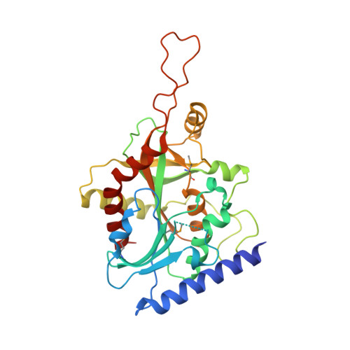
8SWP

Structure of K. lactis PNP bound to hypoxanthine


Experimental Data Snapshot

  • Method: X-RAY DIFFRACTION
  • Resolution: 2.10 Å
  • R-Value Free: 0.236 
  • R-Value Work: 0.199 
  • R-Value Observed: 0.201 

wwPDB Validation   3D Report Full Report


Ligand Structure Quality Assessment 


This is version 1.3 of the entry. See complete history


Literature

Phosphate Binding in PNP Alters Transition-State Analogue Affinity and Subunit Cooperativity.

Minnow, Y.V.T.Schramm, V.L.Almo, S.C.Ghosh, A.

(2023) Biochemistry 62: 3116-3125

  • DOI: https://doi.org/10.1021/acs.biochem.3c00264
  • Primary Citation of Related Structures:  
    8SWP, 8SWQ, 8SWR, 8SWS, 8SWT, 8SWU

  • PubMed Abstract: 

    Purine nucleoside phosphorylases (PNPs) catalyze the phosphorolysis of 6-oxypurine nucleosides with an HPO 4 2- dianion nucleophile. Nucleosides and phosphate occupy distinct pockets in the PNP active site. Evaluation of the HPO 4 2- site by mutagenesis, cooperative binding studies, and thermodynamic and structural analysis demonstrate that alterations in the HPO 4 2- binding site can render PNP inactive and significantly impact subunit cooperativity and binding to transition-state analogue inhibitors. Cooperative interactions between the cationic transition-state analogue and the anionic HPO 4 2- nucleophile demonstrate the importance of reforming the transition-state ensemble for optimal inhibition with transition-state analogues. Altered phosphate binding in the catalytic site mutants helps to explain one of the known lethal PNP deficiency syndromes in humans.


  • Organizational Affiliation

    Department of Biochemistry, Albert Einstein College of Medicine, 1300 Morris Park Avenue, Bronx, New York 10461, United States.


Macromolecules
Find similar proteins by:  (by identity cutoff)  |  3D Structure
Entity ID: 1
MoleculeChains Sequence LengthOrganismDetailsImage
Purine nucleoside phosphorylase
A, B, C, D, E
A, B, C, D, E, F
307Kluyveromyces lactis NRRL Y-1140Mutation(s): 0 
Gene Names: KLLA0_C16621g
EC: 2.4.2.1
UniProt
Find proteins for Q6CSZ6 (Kluyveromyces lactis (strain ATCC 8585 / CBS 2359 / DSM 70799 / NBRC 1267 / NRRL Y-1140 / WM37))
Explore Q6CSZ6 
Go to UniProtKB:  Q6CSZ6
Entity Groups  
Sequence Clusters30% Identity50% Identity70% Identity90% Identity95% Identity100% Identity
UniProt GroupQ6CSZ6
Sequence Annotations
Expand
  • Reference Sequence
Small Molecules
Ligands 2 Unique
IDChains Name / Formula / InChI Key2D Diagram3D Interactions
HPA (Subject of Investigation/LOI)
Query on HPA

Download Ideal Coordinates CCD File 
G [auth A],
I [auth B],
K [auth D],
N [auth E]
HYPOXANTHINE
C5 H4 N4 O
FDGQSTZJBFJUBT-UHFFFAOYSA-N
ACT
Query on ACT

Download Ideal Coordinates CCD File 
H [auth A],
J [auth B],
L [auth D],
M [auth D]
ACETATE ION
C2 H3 O2
QTBSBXVTEAMEQO-UHFFFAOYSA-M
Modified Residues  1 Unique
IDChains TypeFormula2D DiagramParent
MSE
Query on MSE
A, B, C, D, E
A, B, C, D, E, F
L-PEPTIDE LINKINGC5 H11 N O2 SeMET
Experimental Data & Validation

Experimental Data

  • Method: X-RAY DIFFRACTION
  • Resolution: 2.10 Å
  • R-Value Free: 0.236 
  • R-Value Work: 0.199 
  • R-Value Observed: 0.201 
  • Space Group: P 1 21 1
Unit Cell:
Length ( Å )Angle ( ˚ )
a = 89.211α = 90
b = 110.825β = 108.06
c = 97.718γ = 90
Software Package:
Software NamePurpose
PHENIXrefinement
HKL-3000data reduction
SHELXDEphasing
DMphasing
SCALAdata scaling

Structure Validation

View Full Validation Report


Ligand Structure Quality Assessment 


Entry History & Funding Information

Deposition Data


Funding OrganizationLocationGrant Number
National Institutes of Health/National Institute of General Medical Sciences (NIH/NIGMS)United StatesGM041916
National Institutes of Health/National Cancer Institute (NIH/NCI)United StatesCA013330

Revision History  (Full details and data files)

  • Version 1.0: 2023-10-18
    Type: Initial release
  • Version 1.1: 2023-10-25
    Changes: Database references
  • Version 1.2: 2023-11-15
    Changes: Data collection
  • Version 1.3: 2023-11-22
    Changes: Database references