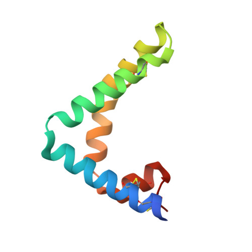
9AXG

Human saposin B in the presence of globotriaosylceramide-NBD


Experimental Data Snapshot

  • Method: X-RAY DIFFRACTION
  • Resolution: 2.68 Å
  • R-Value Free: 0.287 
  • R-Value Work: 0.244 
  • R-Value Observed: 0.248 

Starting Model: experimental
View more details

wwPDB Validation   3D Report Full Report


This is version 1.3 of the entry. See complete history


Literature

Human Saposin B Ligand Binding and Presentation to alpha-Galactosidase A.

Sawyer, T.K.Aral, E.Staros, J.V.Bobst, C.E.Garman, S.C.

(2024) bioRxiv 

  • DOI: https://doi.org/10.1101/2024.04.04.584535
  • Primary Citation of Related Structures:  
    9AVS, 9AXG

  • PubMed Abstract: 

    Sphingolipid activator protein B (saposin B; SapB) is an essential activator of globotriaosylceramide (Gb3) catabolism by α-galactosidase A. However, the manner by which SapB stimulates α-galactosidase A activity remains unknown. To uncover the molecular mechanism of SapB presenting Gb3 to α-galactosidase A, we subjected the fluorescent substrate globotriaosylceramide-nitrobenzoxidazole (Gb3-NBD) to a series of biochemical and structural assays involving SapB. First, we showed that SapB stably binds Gb3-NBD using a fluorescence equilibrium binding assay, isolates Gb3-NBD from micelles, and facilitates α-galactosidase A cleavage of Gb3-NBD in vitro . Second, we crystallized SapB in the presence of Gb3-NBD and validated the ligand-bound assembly. Third, we captured transient interactions between SapB and α-galactosidase A by chemical cross-linking. Finally, we determined the crystal structure of SapB bound to α-galactosidase A. These findings establish general principles for molecular recognition in saposin:hydrolase complexes and highlight the utility of NBD reporter lipids in saposin biochemistry and structural biology.


  • Organizational Affiliation

    Department of Biochemistry & Molecular Biology, Institute of Applied Life Sciences, University of Massachusetts, Amherst, Massachusetts 01003, USA.


Macromolecules
Find similar proteins by:  (by identity cutoff)  |  3D Structure
Entity ID: 1
MoleculeChains Sequence LengthOrganismDetailsImage
Saposin-B
A, B
82Homo sapiensMutation(s): 0 
Gene Names: PSAPGLBASAP1
UniProt & NIH Common Fund Data Resources
Find proteins for P07602 (Homo sapiens)
Explore P07602 
Go to UniProtKB:  P07602
PHAROS:  P07602
GTEx:  ENSG00000197746 
Entity Groups  
Sequence Clusters30% Identity50% Identity70% Identity90% Identity95% Identity100% Identity
UniProt GroupP07602
Sequence Annotations
Expand
  • Reference Sequence
Small Molecules
Ligands 1 Unique
IDChains Name / Formula / InChI Key2D Diagram3D Interactions
MLI
Query on MLI

Download Ideal Coordinates CCD File 
C [auth B]MALONATE ION
C3 H2 O4
OFOBLEOULBTSOW-UHFFFAOYSA-L
Experimental Data & Validation

Experimental Data

  • Method: X-RAY DIFFRACTION
  • Resolution: 2.68 Å
  • R-Value Free: 0.287 
  • R-Value Work: 0.244 
  • R-Value Observed: 0.248 
  • Space Group: P 64
  • Diffraction Data: https://doi.org/10.18430/M39AXG
Unit Cell:
Length ( Å )Angle ( ˚ )
a = 66.908α = 90
b = 66.908β = 90
c = 87.181γ = 120
Software Package:
Software NamePurpose
PHASERphasing
PHENIXrefinement
xia2data reduction
xia2data scaling

Structure Validation

View Full Validation Report


Entry History & Funding Information

Deposition Data


Funding OrganizationLocationGrant Number
National Institutes of Health/National Institute of Diabetes and Digestive and Kidney Disease (NIH/NIDDK)United States5R01DK076877

Revision History  (Full details and data files)

  • Version 1.0: 2024-03-13
    Type: Initial release
  • Version 1.1: 2024-04-17
    Changes: Database references
  • Version 1.2: 2024-04-24
    Changes: Database references
  • Version 1.3: 2024-09-04
    Changes: Database references