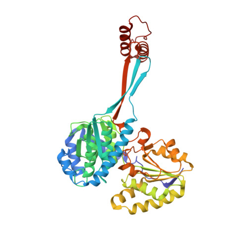
9CP9

Crystal structure of DHPS-3-dehydrogenase, HpsN H319A variant from Cupriavidus pinatubonensis in complex with substrate (R-DHPS) and NADH


Experimental Data Snapshot

  • Method: X-RAY DIFFRACTION
  • Resolution: 2.01 Å
  • R-Value Free: 0.194 
  • R-Value Work: 0.167 

Starting Model: in silico
View more details

wwPDB Validation   3D Report Full Report


Ligand Structure Quality Assessment 


This is version 1.1 of the entry. See complete history


Literature

Structural and kinetic insights into the stereospecific oxidation of R -2,3-dihydroxypropanesulfonate by DHPS-3-dehydrogenase from Cupriavidus pinatubonensis.

Burchill, L.Kaur, A.Nastasovici, A.Lee, M.Williams, S.J.

(2024) Chem Sci 15: 15757-15768

  • DOI: https://doi.org/10.1039/d4sc05114a
  • Primary Citation of Related Structures:  
    8V35, 8V36, 8V37, 9CP7, 9CP8, 9CP9

  • PubMed Abstract: 

    2,3-Dihydroxypropanesulfonate (DHPS) and sulfolactate (SL) are environmentally important organosulfur compounds that play key roles as metabolic currencies in the sulfur cycle. Despite their prevalence, the pathways governing DHPS and SL production remain poorly understood. Here, we study DHPS-3-dehydrogenase from Cupriavidus pinatubonensis ( Cp HpsN), a bacterium capable of utilizing DHPS as a sole carbon source. Kinetic analysis of Cp HpsN reveals a strict preference for R -DHPS, catalyzing its 4-electron oxidation to R -SL, with high specificity for NAD + over NADP + . The 3D structure of Cp HpsN in complex with Zn 2+ , NADH and R -SL, elucidated through X-ray crystallography, reveals a fold akin to bacterial and plant histidinol dehydrogenases with similar coordination geometry around the octahedral Zn 2+ centre and involving the sulfonate group as a ligand. A key residue, His126, distinguishes DHPS dehydrogenases from histidinol dehydrogenases, by structural recognition of the sulfonate substrate of the former. Site-directed mutagenesis pinpoints Glu318, His319, and Asp352 as active-site residues important for the catalytic activity of Cp HpsN. Taxonomic and pathway distribution analysis reveals the prevalence of HpsN homologues within different pathways of DHPS catabolism and across bacterial classes including Alpha-, Beta-, Gamma-, and Deltaproteobacteria and Desulfobacteria, emphasizing its importance in the biogeochemical sulfur cycle.


  • Organizational Affiliation

    School of Chemistry, Bio21 Molecular Science and Biotechnology Institute, University of Melbourne Parkville Victoria 3010 Australia [email protected] [email protected].


Macromolecules
Find similar proteins by:  (by identity cutoff)  |  3D Structure
Entity ID: 1
MoleculeChains Sequence LengthOrganismDetailsImage
Sulfopropanediol 3-dehydrogenase
A, B
437Cupriavidus pinatubonensis JMP134Mutation(s): 1 
Gene Names: hpsNReut_C6092
EC: 1.1.1.308
UniProt
Find proteins for Q46N53 (Cupriavidus pinatubonensis (strain JMP 134 / LMG 1197))
Explore Q46N53 
Go to UniProtKB:  Q46N53
Entity Groups  
Sequence Clusters30% Identity50% Identity70% Identity90% Identity95% Identity100% Identity
UniProt GroupQ46N53
Sequence Annotations
Expand
  • Reference Sequence
Small Molecules
Ligands 4 Unique
IDChains Name / Formula / InChI Key2D Diagram3D Interactions
NAI (Subject of Investigation/LOI)
Query on NAI

Download Ideal Coordinates CCD File 
D [auth A],
I [auth B]
1,4-DIHYDRONICOTINAMIDE ADENINE DINUCLEOTIDE
C21 H29 N7 O14 P2
BOPGDPNILDQYTO-NNYOXOHSSA-N
A1AZH (Subject of Investigation/LOI)
Query on A1AZH

Download Ideal Coordinates CCD File 
E [auth A],
J [auth B]
(2R)-2,3-dihydroxypropane-1-sulfonic acid
C3 H8 O5 S
YPFUJZAAZJXMIP-GSVOUGTGSA-N
SO4
Query on SO4

Download Ideal Coordinates CCD File 
F [auth A],
G [auth A],
K [auth B]
SULFATE ION
O4 S
QAOWNCQODCNURD-UHFFFAOYSA-L
ZN (Subject of Investigation/LOI)
Query on ZN

Download Ideal Coordinates CCD File 
C [auth A],
H [auth B]
ZINC ION
Zn
PTFCDOFLOPIGGS-UHFFFAOYSA-N
Experimental Data & Validation

Experimental Data

  • Method: X-RAY DIFFRACTION
  • Resolution: 2.01 Å
  • R-Value Free: 0.194 
  • R-Value Work: 0.167 
  • Space Group: P 1 21 1
Unit Cell:
Length ( Å )Angle ( ˚ )
a = 83.324α = 90
b = 75.809β = 117.099
c = 84.674γ = 90
Software Package:
Software NamePurpose
REFMACrefinement
XDSdata reduction
Aimlessdata scaling
PHASERphasing

Structure Validation

View Full Validation Report


Ligand Structure Quality Assessment 


Entry History & Funding Information

Deposition Data

  • Released Date: 2024-09-25 
  • Deposition Author(s): Lee, M.

Funding OrganizationLocationGrant Number
Australian Research Council (ARC)Australia--

Revision History  (Full details and data files)

  • Version 1.0: 2024-09-25
    Type: Initial release
  • Version 1.1: 2024-10-09
    Changes: Database references, Structure summary