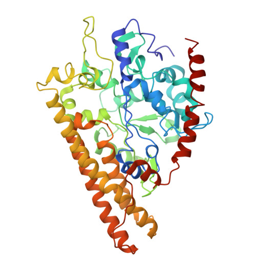
9CUF

Room temperature SSX structure of ccNiR


Experimental Data Snapshot

  • Method: X-RAY DIFFRACTION
  • Resolution: 3.30 Å
  • R-Value Free: 0.298 
  • R-Value Work: 0.212 
  • R-Value Observed: 0.221 

Starting Model: experimental
View more details

wwPDB Validation   3D Report Full Report


Ligand Structure Quality Assessment 


This is version 1.1 of the entry. See complete history


Literature

Exploiting fourth-generation synchrotron radiation for enzyme and photoreceptor characterization.

Malla, T.N.Muniyappan, S.Menendez, D.Ogukwe, F.Dale, A.N.Clayton, J.D.Weatherall, D.D.Karki, P.Dangi, S.Mandella, V.Pacheco, A.A.Stojkovic, E.A.Rose, S.L.Orlans, J.Basu, S.de Sanctis, D.Schmidt, M.

(2025) IUCrJ 12: 36-48

  • DOI: https://doi.org/10.1107/S2052252524010868
  • Primary Citation of Related Structures:  
    9CUF, 9D10, 9D2H

  • PubMed Abstract: 

    The upgrade of the European Synchrotron Radiation Facility (ESRF) in Grenoble, France to an Extremely Brilliant Source (EBS) is expected to enable time-resolved synchrotron serial crystallography (SSX) experiments with sub-millisecond time resolution. ID29 is a new beamline dedicated to SSX experiments at ESRF-EBS. Here, we report experiments emerging from the initial phase of user operation at ID29. We first used microcrystals of photoactive yellow protein as a model system to exploit the potential of microsecond pulses for SSX. Subsequently, we investigated microcrystals of cytochrome c nitrite reductase (ccNiR) with microsecond X-ray pulses. CcNiR is a decaheme protein that is ideal for the investigation of radiation damage at the various heme-iron sites. Finally, we performed a proof-of-concept subsecond time-resolved SSX experiment by photoactivating microcrystals of a myxobacterial phytochrome.


  • Organizational Affiliation

    Department of Physics, University of Wisconsin-Milwaukee, Milwaukee, USA.


Macromolecules
Find similar proteins by:  (by identity cutoff)  |  3D Structure
Entity ID: 1
MoleculeChains Sequence LengthOrganismDetailsImage
Cytochrome c-552
A, B
439Shewanella oneidensisMutation(s): 0 
Gene Names: nrfASO_3980
EC: 1.7.2.2
UniProt
Find proteins for Q8EAC7 (Shewanella oneidensis (strain ATCC 700550 / JCM 31522 / CIP 106686 / LMG 19005 / NCIMB 14063 / MR-1))
Explore Q8EAC7 
Go to UniProtKB:  Q8EAC7
Entity Groups  
Sequence Clusters30% Identity50% Identity70% Identity90% Identity95% Identity100% Identity
UniProt GroupQ8EAC7
Sequence Annotations
Expand
  • Reference Sequence
Small Molecules
Ligands 3 Unique
IDChains Name / Formula / InChI Key2D Diagram3D Interactions
PE3
Query on PE3

Download Ideal Coordinates CCD File 
O [auth B]3,6,9,12,15,18,21,24,27,30,33,36,39-TRIDECAOXAHENTETRACONTANE-1,41-DIOL
C28 H58 O15
ILLKMACMBHTSHP-UHFFFAOYSA-N
HEC (Subject of Investigation/LOI)
Query on HEC

Download Ideal Coordinates CCD File 
C [auth A]
D [auth A]
E [auth A]
F [auth A]
G [auth A]
C [auth A],
D [auth A],
E [auth A],
F [auth A],
G [auth A],
I [auth B],
J [auth B],
K [auth B],
L [auth B],
M [auth B]
HEME C
C34 H34 Fe N4 O4
HXQIYSLZKNYNMH-LJNAALQVSA-N
CA
Query on CA

Download Ideal Coordinates CCD File 
H [auth A],
N [auth B]
CALCIUM ION
Ca
BHPQYMZQTOCNFJ-UHFFFAOYSA-N
Experimental Data & Validation

Experimental Data

  • Method: X-RAY DIFFRACTION
  • Resolution: 3.30 Å
  • R-Value Free: 0.298 
  • R-Value Work: 0.212 
  • R-Value Observed: 0.221 
  • Space Group: P 21 21 21
Unit Cell:
Length ( Å )Angle ( ˚ )
a = 50.84α = 90
b = 95.47β = 90
c = 228.2γ = 90
Software Package:
Software NamePurpose
PHENIXrefinement
CrystFELdata scaling
PHASERphasing
CrystFELdata reduction

Structure Validation

View Full Validation Report


Ligand Structure Quality Assessment 


Entry History & Funding Information

Deposition Data


Funding OrganizationLocationGrant Number
National Science Foundation (NSF, United States)United States--

Revision History  (Full details and data files)

  • Version 1.0: 2025-01-15
    Type: Initial release
  • Version 1.1: 2025-01-22
    Changes: Database references