


Crystal Structure of 5'-Deoxy-5'-methylthioadenosine phosphorylase from Aeropyrum pernix complex with 5'-Deoxy-5'-methylthioadenosine 353K
Iizuka, Y., Kikuchi, M., Yamauchi, T., Tsunoda, M.To be published.
Experimental Data Snapshot
Starting Model: experimental
View more details
Entity ID: 1 | |||||
|---|---|---|---|---|---|
| Molecule | Chains | Sequence Length | Organism | Details | Image |
| S-methyl-5'-thioadenosine phosphorylase | 275 | Aeropyrum pernix K1 | Mutation(s): 0 Gene Names: mtnP, APE_1885 EC: 2.4.2.28 | 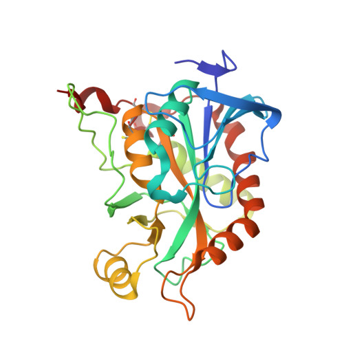 | |
UniProt | |||||
Find proteins for Q9YAQ8 (Aeropyrum pernix (strain ATCC 700893 / DSM 11879 / JCM 9820 / NBRC 100138 / K1)) Explore Q9YAQ8 Go to UniProtKB: Q9YAQ8 | |||||
Entity Groups | |||||
| Sequence Clusters | 30% Identity50% Identity70% Identity90% Identity95% Identity100% Identity | ||||
| UniProt Group | Q9YAQ8 | ||||
Sequence AnnotationsExpand | |||||
| |||||
| Ligands 2 Unique | |||||
|---|---|---|---|---|---|
| ID | Chains | Name / Formula / InChI Key | 2D Diagram | 3D Interactions | |
| MTA (Subject of Investigation/LOI) Query on MTA | C [auth A] | 5'-DEOXY-5'-METHYLTHIOADENOSINE C11 H15 N5 O3 S WUUGFSXJNOTRMR-IOSLPCCCSA-N |  | ||
| PO4 (Subject of Investigation/LOI) Query on PO4 | B [auth A] | PHOSPHATE ION O4 P NBIIXXVUZAFLBC-UHFFFAOYSA-K |  | ||
| Length ( Å ) | Angle ( ˚ ) |
|---|---|
| a = 79.048 | α = 90 |
| b = 79.048 | β = 90 |
| c = 233.48 | γ = 120 |
| Software Name | Purpose |
|---|---|
| REFMAC | refinement |
| Coot | model building |
| MOLREP | phasing |
| Aimless | data scaling |
| XDS | data reduction |
| Funding Organization | Location | Grant Number |
|---|---|---|
| Not funded | -- |
RCSB PDB is hosted by
RCSB PDB is a member of the

