1DVG

CRYSTAL STRUCTURE OF RAT HEME OXYGENASE-1 IN COMPLEX WITH HEME; SELELENO-METHIONINE DERIVATIVE, MUTATED AT M51T,M93L,M155L,M191L.


Experimental Data Snapshot

  • Method: X-RAY DIFFRACTION
  • Resolution: 2.20 Å
  • R-Value Free: 0.258 
  • R-Value Work: 0.212 

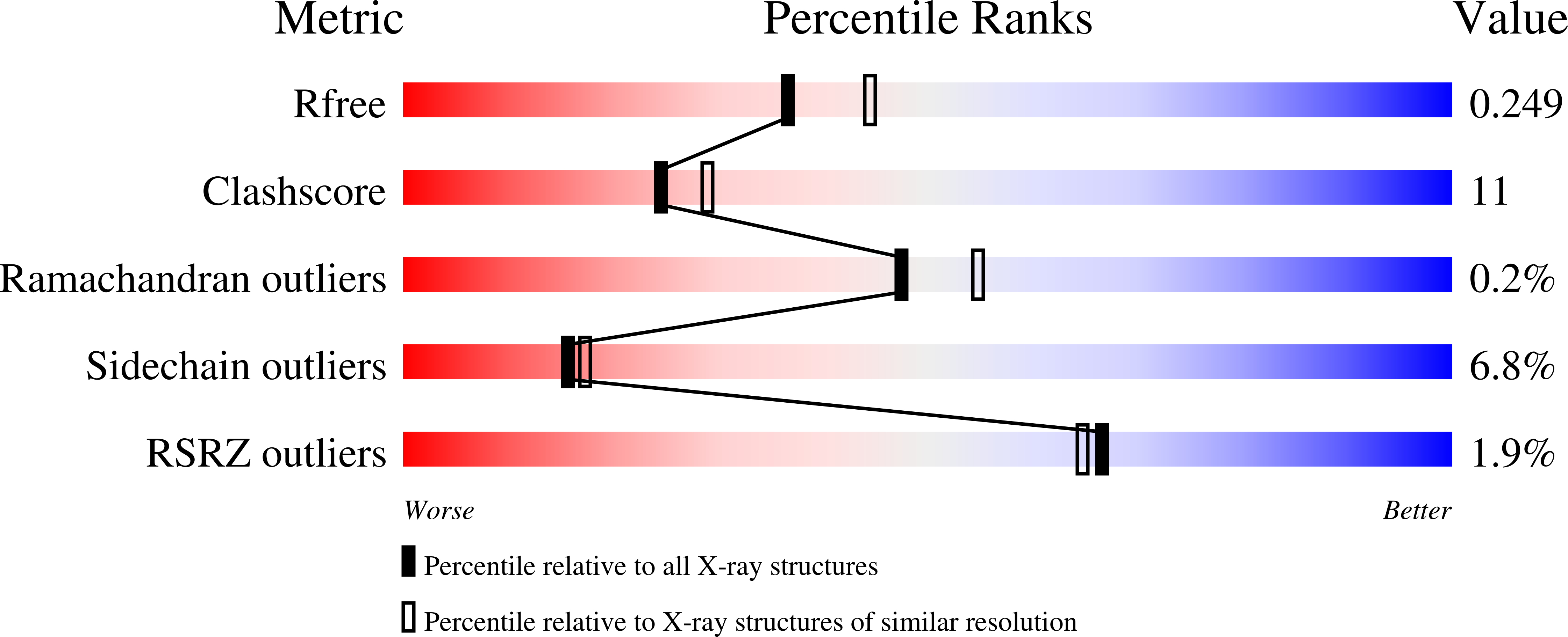
wwPDB Validation   3D Report Full Report


Ligand Structure Quality Assessment 


This is version 1.4 of the entry. See complete history


Literature

Crystal structure of rat heme oxygenase-1 in complex with heme.

Sugishima, M.Omata, Y.Kakuta, Y.Sakamoto, H.Noguchi, M.Fukuyama, K.

(2000) FEBS Lett 471: 61-66

  • DOI: https://doi.org/10.1016/s0014-5793(00)01353-3
  • Primary Citation of Related Structures:  
    1DVE, 1DVG

  • PubMed Abstract: 

    Heme oxygenase catalyzes the oxidative cleavage of protoheme to biliverdin, the first step of heme metabolism utilizing O(2) and NADPH. We determined the crystal structures of rat heme oxygenase-1 (HO-1)-heme and selenomethionyl HO-1-heme complexes. Heme is sandwiched between two helices with the delta-meso edge of the heme being exposed to the surface. Gly143N forms a hydrogen bond to the distal ligand of heme, OH(-). The distance between Gly143N and the ligand is shorter than that in the human HO-1-heme complex. This difference may be related to a pH-dependent change of the distal ligand of heme. Flexibility of the distal helix may control the stability of the coordination of the distal ligand to heme iron. The possible role of Gly143 in the heme oxygenase reaction is discussed.


  • Organizational Affiliation

    Department of Biology, Graduate School of Science, Osaka University, Toyonaka, Osaka, Japan.


Macromolecules
Find similar proteins by:  (by identity cutoff)  |  3D Structure
Entity ID: 1
MoleculeChains Sequence LengthOrganismDetailsImage
HEME OXYGENASE-1
A, B
267Rattus norvegicusMutation(s): 8 
EC: 1.14.99.3 (PDB Primary Data), 1.14.14.18 (UniProt)
UniProt
Find proteins for P06762 (Rattus norvegicus)
Explore P06762 
Go to UniProtKB:  P06762
Entity Groups  
Sequence Clusters30% Identity50% Identity70% Identity90% Identity95% Identity100% Identity
UniProt GroupP06762
Sequence Annotations
Expand
  • Reference Sequence
Small Molecules
Modified Residues  1 Unique
IDChains TypeFormula2D DiagramParent
MSE
Query on MSE
A, B
L-PEPTIDE LINKINGC5 H11 N O2 SeMET
Experimental Data & Validation

Experimental Data

  • Method: X-RAY DIFFRACTION
  • Resolution: 2.20 Å
  • R-Value Free: 0.258 
  • R-Value Work: 0.212 
  • Space Group: P 43
Unit Cell:
Length ( Å )Angle ( ˚ )
a = 54.853α = 90
b = 54.853β = 90
c = 187.558γ = 90
Software Package:
Software NamePurpose
CNSrefinement
DENZOdata reduction
SCALEPACKdata scaling
CNSphasing

Structure Validation

View Full Validation Report


Ligand Structure Quality Assessment 


Entry History 

Deposition Data

Revision History  (Full details and data files)

  • Version 1.0: 2000-04-12
    Type: Initial release
  • Version 1.1: 2008-04-27
    Changes: Version format compliance
  • Version 1.2: 2011-07-13
    Changes: Version format compliance
  • Version 1.3: 2021-11-03
    Changes: Database references, Derived calculations
  • Version 1.4: 2024-10-30
    Changes: Data collection, Structure summary