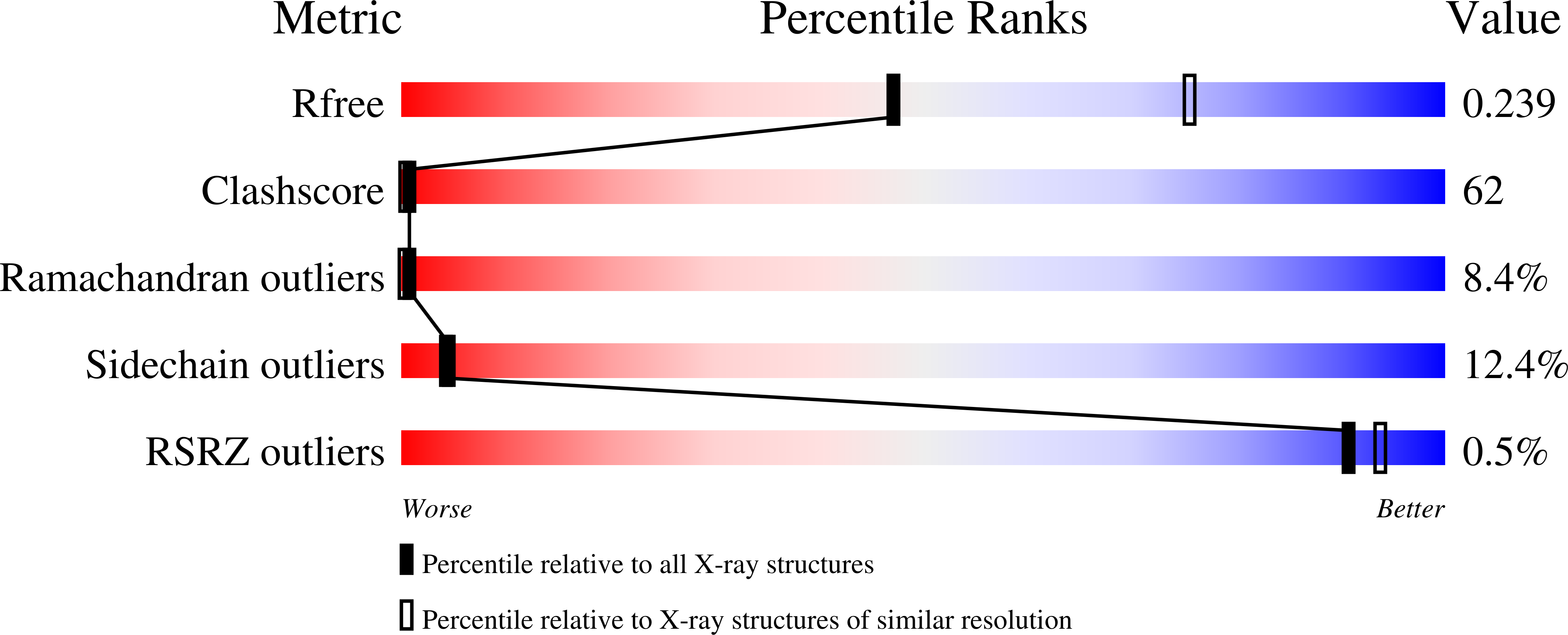
Crystal structure of rat apo-heme oxygenase-1 (HO-1): mechanism of heme binding in HO-1 inferred from structural comparison of the apo and heme complex forms
Sugishima, M., Sakamoto, H., Kakuta, Y., Omata, Y., Hayashi, S., Noguchi, M., Fukuyama, K.(2002) Biochemistry 41: 7293-7300
- PubMed: 12044160
- DOI: https://doi.org/10.1021/bi025662a
- Primary Citation of Related Structures:
1IRM - PubMed Abstract:
Heme oxygenase (HO) catalyzes the oxidative cleavage of heme to biliverdin by utilizing O(2) and NADPH. HO (apoHO) was crystallized as twinned P3(2) with three molecules per asymmetric unit, and its crystal structure was determined at 2.55 A resolution. Structural comparison of apoHO and its complex with heme (HO-heme) showed three distinct differences. First, the A helix of the eight alpha-helices (A-H) in HO-heme, which includes the proximal ligand of heme (His25), is invisible in apoHO. In addition, the B helix, a portion of which builds the heme pocket, is shifted toward the heme pocket in apoHO. Second, Gln38 is shifted toward the position where the alpha-meso carbon of heme is located in HO-heme. Nepsilon of Gln38 is hydrogen-bonded to the carbonyl group of Glu29 located at the C-terminal side of the A helix in HO-heme, indicative that this hydrogen bond restrains the angle between the A and B helices in HO-heme. Third, the amide group of Gly143 in the F helix is directed outward from the heme pocket in apoHO, whereas it is directed toward the distal ligand of heme in HO-heme. This means that the F helix around Gly143 must change its conformation to accommodate heme binding. The apoHO structure has the characteristic that the helix on one side of the heme pocket fluctuates, whereas the rest of the structure is similar to that of HO-heme, as observed in such hemoproteins as myoglobin and cytochromes b(5) and b(562). These structural features of apoHO suggest that the orientation of the proximal helix and the position of His25 are fixed upon heme binding.
Organizational Affiliation:
Department of Biology, Graduate School of Science, Osaka University, Toyonaka, Osaka 560-0043, Japan.


