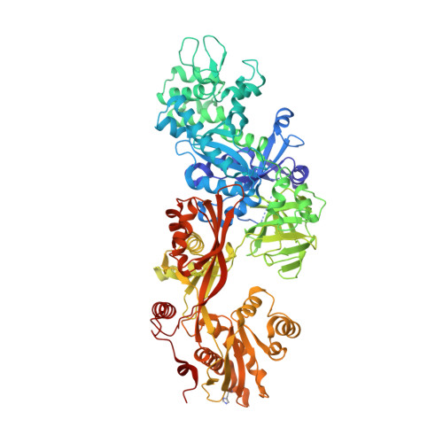
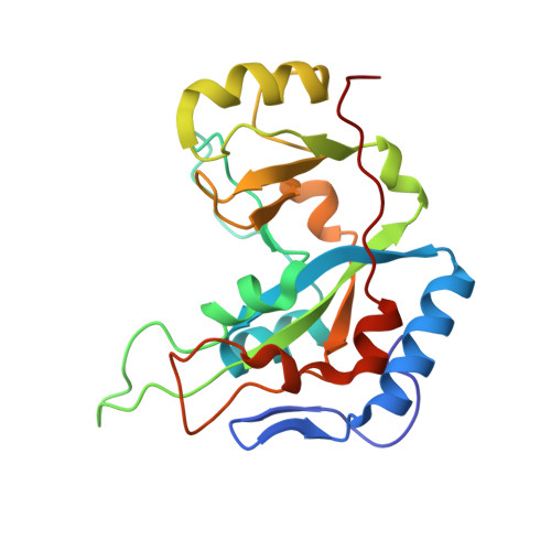
1ZM3

Structure of the apo eEF2-ETA complex


Experimental Data Snapshot

  • Method: X-RAY DIFFRACTION
  • Resolution: 3.07 Å
  • R-Value Free: 0.282 
  • R-Value Work: 0.249 
  • R-Value Observed: 0.250 

Starting Models: experimental
View more details

wwPDB Validation   3D Report Full Report


This is version 1.3 of the entry. See complete history


Literature

Exotoxin A-eEF2 complex structure indicates ADP ribosylation by ribosome mimicry.

Joergensen, R.Merrill, A.R.Yates, S.P.Marquez, V.E.Schwan, A.L.Boesen, T.Andersen, G.R.

(2005) Nature 436: 979-984

  • DOI: https://doi.org/10.1038/nature03871
  • Primary Citation of Related Structures:  
    1ZM2, 1ZM3, 1ZM4, 1ZM9

  • PubMed Abstract: 

    The bacteria causing diphtheria, whooping cough, cholera and other diseases secrete mono-ADP-ribosylating toxins that modify intracellular proteins. Here, we describe four structures of a catalytically active complex between a fragment of Pseudomonas aeruginosa exotoxin A (ETA) and its protein substrate, translation elongation factor 2 (eEF2). The target residue in eEF2, diphthamide (a modified histidine), spans across a cleft and faces the two phosphates and a ribose of the non-hydrolysable NAD+ analogue, betaTAD. This suggests that the diphthamide is involved in triggering NAD+ cleavage and interacting with the proposed oxacarbenium intermediate during the nucleophilic substitution reaction, explaining the requirement of diphthamide for ADP ribosylation. Diphtheria toxin may recognize eEF2 in a manner similar to ETA. Notably, the toxin-bound betaTAD phosphates mimic the phosphate backbone of two nucleotides in a conformational switch of 18S rRNA, thereby achieving universal recognition of eEF2 by ETA.


  • Organizational Affiliation

    Centre for Structural Biology, Department of Molecular Biology, University of Aarhus, Gustav Wieds Vej 10C, DK-8000, Denmark.


Macromolecules
Find similar proteins by:  (by identity cutoff)  |  3D Structure
Entity ID: 1
MoleculeChains Sequence LengthOrganismDetailsImage
Elongation factor 2
A, C, E
842Saccharomyces cerevisiaeMutation(s): 1 
EC: 3.6.5
UniProt
Find proteins for P32324 (Saccharomyces cerevisiae (strain ATCC 204508 / S288c))
Explore P32324 
Go to UniProtKB:  P32324
Entity Groups  
Sequence Clusters30% Identity50% Identity70% Identity90% Identity95% Identity100% Identity
UniProt GroupP32324
Sequence Annotations
Expand
  • Reference Sequence
Find similar proteins by:  (by identity cutoff)  |  3D Structure
Entity ID: 2
MoleculeChains Sequence LengthOrganismDetailsImage
exotoxin A
B, D, F
207Pseudomonas aeruginosaMutation(s): 0 
EC: 2.4.2.36
UniProt
Find proteins for P11439 (Pseudomonas aeruginosa (strain ATCC 15692 / DSM 22644 / CIP 104116 / JCM 14847 / LMG 12228 / 1C / PRS 101 / PAO1))
Explore P11439 
Go to UniProtKB:  P11439
Entity Groups  
Sequence Clusters30% Identity50% Identity70% Identity90% Identity95% Identity100% Identity
UniProt GroupP11439
Sequence Annotations
Expand
  • Reference Sequence
Small Molecules
Modified Residues  1 Unique
IDChains TypeFormula2D DiagramParent
DDE
Query on DDE
A, C, E
L-PEPTIDE LINKINGC13 H24 N5 O3HIS
Experimental Data & Validation

Experimental Data

  • Method: X-RAY DIFFRACTION
  • Resolution: 3.07 Å
  • R-Value Free: 0.282 
  • R-Value Work: 0.249 
  • R-Value Observed: 0.250 
  • Space Group: C 1 2 1
Unit Cell:
Length ( Å )Angle ( ˚ )
a = 329.43α = 90
b = 69.09β = 103.46
c = 190.8γ = 90
Software Package:
Software NamePurpose
XDSdata scaling
XDSdata reduction
MOLREPphasing
CNSrefinement

Structure Validation

View Full Validation Report


Entry History 

Deposition Data

Revision History  (Full details and data files)

  • Version 1.0: 2005-05-24
    Type: Initial release
  • Version 1.1: 2008-04-30
    Changes: Version format compliance
  • Version 1.2: 2011-07-13
    Changes: Version format compliance
  • Version 1.3: 2023-08-23
    Changes: Data collection, Database references, Derived calculations, Refinement description