2BMQ

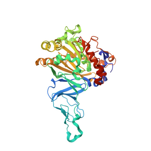
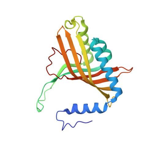
The Crystal Structure of Nitrobenzene Dioxygenase in complex with nitrobenzene


Experimental Data Snapshot

  • Method: X-RAY DIFFRACTION
  • Resolution: 1.55 Å
  • R-Value Free: 0.192 
  • R-Value Work: 0.167 
  • R-Value Observed: 0.169 

Starting Model: experimental
View more details

wwPDB Validation   3D Report Full Report


This is version 1.5 of the entry. See complete history


Literature

Structural insight into the dioxygenation of nitroarene compounds: the crystal structure of nitrobenzene dioxygenase.

Friemann, R.Ivkovic-Jensen, M.M.Lessner, D.J.Yu, C.L.Gibson, D.T.Parales, R.E.Eklund, H.Ramaswamy, S.

(2005) J Mol Biol 348: 1139-1151

  • DOI: https://doi.org/10.1016/j.jmb.2005.03.052
  • Primary Citation of Related Structures:  
    2BMO, 2BMQ, 2BMR

  • PubMed Abstract: 

    Nitroaromatic compounds are used extensively in many industrial processes and have been released into the environment where they are considered environmental pollutants. Nitroaromatic compounds, in general, are resistant to oxidative attack due to the electron-withdrawing nature of the nitro groups and the stability of the benzene ring. However, the bacterium Comamonas sp. strain JS765 can grow with nitrobenzene as a sole source of carbon, nitrogen and energy. Biodegradation is initiated by the nitrobenzene dioxygenase (NBDO) system. We have determined the structure of NBDO, which has a hetero-hexameric structure similar to that of several other Rieske non-heme iron dioxygenases. The catalytic subunit contains a Rieske iron-sulfur center and an active-site mononuclear iron atom. The structures of complexes with substrates nitrobenzene and 3-nitrotoluene reveal the structural basis for its activity with nitroarenes. The substrate pocket contains an asparagine residue that forms a hydrogen bond to the nitro-group of the substrate, and orients the substrate in relation to the active-site mononuclear iron atom, positioning the molecule for oxidation at the nitro-substituted carbon.


  • Organizational Affiliation

    Department of Molecular Biology, Swedish University of Agricultural Sciences, Uppsala Biomedical Center, Box 590 S-751 24 Uppsala, Sweden. [email protected]


Macromolecules
Find similar proteins by:  (by identity cutoff)  |  3D Structure
Entity ID: 1
MoleculeChains Sequence LengthOrganismDetailsImage
OXYGENASE-ALPHA NBDO447Comamonas sp. JS765Mutation(s): 0 
UniProt
Find proteins for Q8RTL4 (Comamonas sp. JS765)
Explore Q8RTL4 
Go to UniProtKB:  Q8RTL4
Entity Groups  
Sequence Clusters30% Identity50% Identity70% Identity90% Identity95% Identity100% Identity
UniProt GroupQ8RTL4
Sequence Annotations
Expand
  • Reference Sequence
Find similar proteins by:  (by identity cutoff)  |  3D Structure
Entity ID: 2
MoleculeChains Sequence LengthOrganismDetailsImage
OXYGENASE-BETA NBDO194Comamonas sp. JS765Mutation(s): 0 
UniProt
Find proteins for Q8RTL3 (Comamonas sp. JS765)
Explore Q8RTL3 
Go to UniProtKB:  Q8RTL3
Entity Groups  
Sequence Clusters30% Identity50% Identity70% Identity90% Identity95% Identity100% Identity
UniProt GroupQ8RTL3
Sequence Annotations
Expand
  • Reference Sequence
Small Molecules
Ligands 6 Unique
IDChains Name / Formula / InChI Key2D Diagram3D Interactions
FES
Query on FES

Download Ideal Coordinates CCD File 
C [auth A]FE2/S2 (INORGANIC) CLUSTER
Fe2 S2
NIXDOXVAJZFRNF-UHFFFAOYSA-N
NBZ
Query on NBZ

Download Ideal Coordinates CCD File 
E [auth A]NITROBENZENE
C6 H5 N O2
LQNUZADURLCDLV-UHFFFAOYSA-N
EDO
Query on EDO

Download Ideal Coordinates CCD File 
F [auth A]
G [auth A]
N [auth B]
O [auth B]
P [auth B]
F [auth A],
G [auth A],
N [auth B],
O [auth B],
P [auth B],
Q [auth B]
1,2-ETHANEDIOL
C2 H6 O2
LYCAIKOWRPUZTN-UHFFFAOYSA-N
NI
Query on NI

Download Ideal Coordinates CCD File 
L [auth B],
M [auth B]
NICKEL (II) ION
Ni
VEQPNABPJHWNSG-UHFFFAOYSA-N
FE
Query on FE

Download Ideal Coordinates CCD File 
D [auth A]FE (III) ION
Fe
VTLYFUHAOXGGBS-UHFFFAOYSA-N
EOH
Query on EOH

Download Ideal Coordinates CCD File 
H [auth A]
I [auth A]
J [auth A]
K [auth A]
R [auth B]
H [auth A],
I [auth A],
J [auth A],
K [auth A],
R [auth B],
S [auth B]
ETHANOL
C2 H6 O
LFQSCWFLJHTTHZ-UHFFFAOYSA-N
Experimental Data & Validation

Experimental Data

  • Method: X-RAY DIFFRACTION
  • Resolution: 1.55 Å
  • R-Value Free: 0.192 
  • R-Value Work: 0.167 
  • R-Value Observed: 0.169 
  • Space Group: P 63
Unit Cell:
Length ( Å )Angle ( ˚ )
a = 121.533α = 90
b = 121.533β = 90
c = 84.179γ = 120
Software Package:
Software NamePurpose
REFMACrefinement
MOSFLMdata reduction
SCALAdata scaling

Structure Validation

View Full Validation Report


Entry History 

Deposition Data

Revision History  (Full details and data files)

  • Version 1.0: 2005-05-04
    Type: Initial release
  • Version 1.1: 2011-05-08
    Changes: Version format compliance
  • Version 1.2: 2011-07-13
    Changes: Version format compliance
  • Version 1.3: 2018-01-17
    Changes: Advisory, Data collection
  • Version 1.4: 2018-02-28
    Changes: Database references, Source and taxonomy
  • Version 1.5: 2023-12-13
    Changes: Advisory, Data collection, Database references, Derived calculations, Other, Refinement description