2I2W

Crystal Structure of Escherichia Coli Phosphoheptose Isomerase


Experimental Data Snapshot

  • Method: X-RAY DIFFRACTION
  • Resolution: 1.95 Å
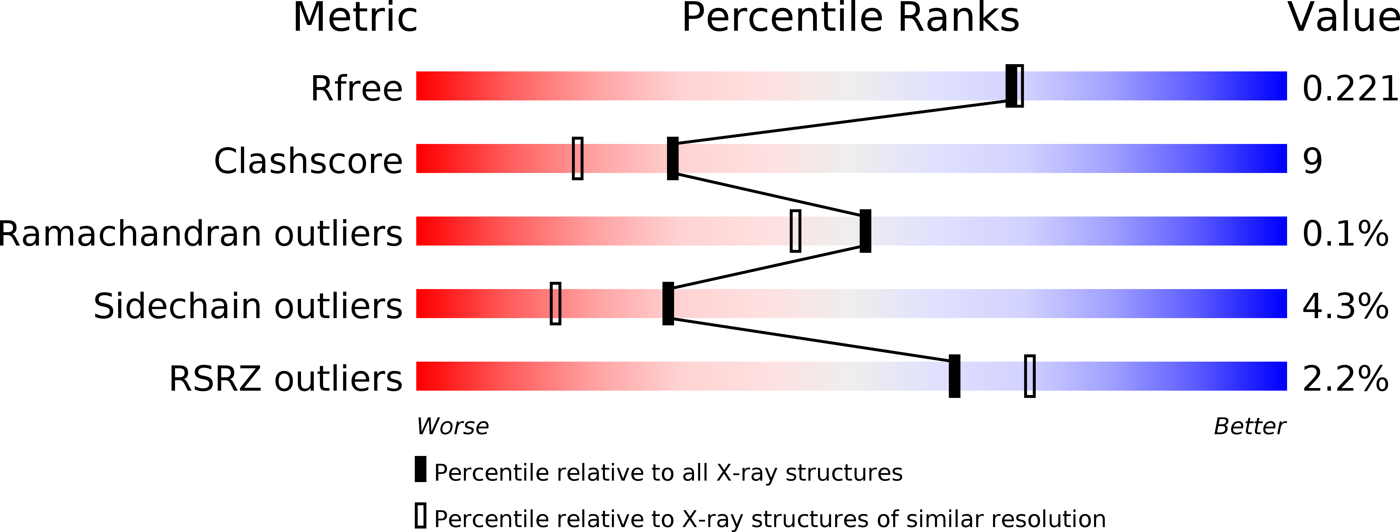
  • R-Value Free: 0.219 
  • R-Value Work: 0.170 
  • R-Value Observed: 0.172 

wwPDB Validation   3D Report Full Report


This is version 1.1 of the entry. See complete history


Literature

Structure and Function of Sedoheptulose-7-phosphate Isomerase, a Critical Enzyme for Lipopolysaccharide Biosynthesis and a Target for Antibiotic Adjuvants

Taylor, P.L.Blakely, K.M.de Leon, G.P.Walker, J.R.McArthur, F.Evdokimova, E.Zhang, K.Valvano, M.A.Wright, G.D.Junop, M.S.

(2008) J Biol Chem 283: 2835-2845

  • DOI: https://doi.org/10.1074/jbc.M706163200
  • Primary Citation of Related Structures:  
    1X92, 2I22, 2I2W, 3BJZ

  • PubMed Abstract: 

    The barrier imposed by lipopolysaccharide (LPS) in the outer membrane of Gram-negative bacteria presents a significant challenge in treatment of these organisms with otherwise effective hydrophobic antibiotics. The absence of L-glycero-D-manno-heptose in the LPS molecule is associated with a dramatically increased bacterial susceptibility to hydrophobic antibiotics and thus enzymes in the ADP-heptose biosynthesis pathway are of significant interest. GmhA catalyzes the isomerization of D-sedoheptulose 7-phosphate into D-glycero-D-manno-heptose 7-phosphate, the first committed step in the formation of ADP-heptose. Here we report structures of GmhA from Escherichia coli and Pseudomonas aeruginosa in apo, substrate, and product-bound forms, which together suggest that GmhA adopts two distinct conformations during isomerization through reorganization of quaternary structure. Biochemical characterization of GmhA mutants, combined with in vivo analysis of LPS biosynthesis and novobiocin susceptibility, identifies key catalytic residues. We postulate GmhA acts through an enediol-intermediate isomerase mechanism.


  • Organizational Affiliation

    Department of Biochemistry and Biomedical Sciences, DeGroote School of Medicine, McMaster University, 1200 Main Street West, Hamilton, Ontario, Canada.


Macromolecules
Find similar proteins by:  (by identity cutoff)  |  3D Structure
Entity ID: 1
MoleculeChains Sequence LengthOrganismDetailsImage
Phosphoheptose isomerase
A, B, C, D
212Escherichia coliMutation(s): 0 
Gene Names: gmhAlpcAtfrAb0222
EC: 5.3.1 (PDB Primary Data), 5.3.1.28 (UniProt)
UniProt
Find proteins for P63224 (Escherichia coli (strain K12))
Explore P63224 
Go to UniProtKB:  P63224
Entity Groups  
Sequence Clusters30% Identity50% Identity70% Identity90% Identity95% Identity100% Identity
UniProt GroupP63224
Sequence Annotations
Expand
  • Reference Sequence
Small Molecules
Ligands 1 Unique
IDChains Name / Formula / InChI Key2D Diagram3D Interactions
GOL
Query on GOL

Download Ideal Coordinates CCD File 
E [auth D]GLYCEROL
C3 H8 O3
PEDCQBHIVMGVHV-UHFFFAOYSA-N
Experimental Data & Validation

Experimental Data

  • Method: X-RAY DIFFRACTION
  • Resolution: 1.95 Å
  • R-Value Free: 0.219 
  • R-Value Work: 0.170 
  • R-Value Observed: 0.172 
  • Space Group: P 21 21 21
Unit Cell:
Length ( Å )Angle ( ˚ )
a = 83.93α = 90
b = 89.61β = 90
c = 106.91γ = 90
Software Package:
Software NamePurpose
REFMACrefinement
CrystalCleardata collection
CrystalCleardata reduction
CrystalCleardata scaling
MOLREPphasing

Structure Validation

View Full Validation Report


Entry History 

Deposition Data

Revision History  (Full details and data files)

  • Version 1.0: 2007-08-21
    Type: Initial release
  • Version 1.1: 2011-07-13
    Changes: Derived calculations, Version format compliance