2II3

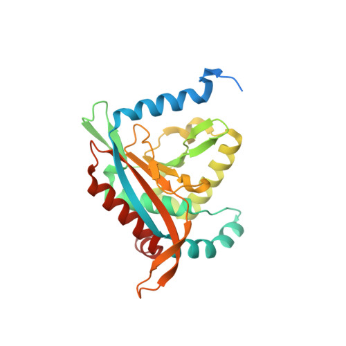
Crystal structure of a cubic core of the dihydrolipoamide acyltransferase (E2b) component in the branched-chain alpha-ketoacid dehydrogenase complex (BCKDC), Oxidized Coenzyme A-bound form


Experimental Data Snapshot

  • Method: X-RAY DIFFRACTION
  • Resolution: 2.17 Å
  • R-Value Free: 0.259 
  • R-Value Work: 0.202 
  • R-Value Observed: 0.205 

Starting Model: experimental
View more details

wwPDB Validation   3D Report Full Report


Ligand Structure Quality Assessment 


This is version 1.4 of the entry. See complete history


Literature

A synchronized substrate-gating mechanism revealed by cubic-core structure of the bovine branched-chain alpha-ketoacid dehydrogenase complex.

Kato, M.Wynn, R.M.Chuang, J.L.Brautigam, C.A.Custorio, M.Chuang, D.T.

(2006) EMBO J 25: 5983-5994

  • DOI: https://doi.org/10.1038/sj.emboj.7601444
  • Primary Citation of Related Structures:  
    2IHW, 2II3, 2II4, 2II5

  • PubMed Abstract: 

    The dihydrolipoamide acyltransferase (E2b) component of the branched-chain alpha-ketoacid dehydrogenase complex forms a cubic scaffold that catalyzes acyltransfer from S-acyldihydrolipoamide to CoA to produce acyl-CoA. We have determined the first crystal structures of a mammalian (bovine) E2b core domain with and without a bound CoA or acyl-CoA. These structures reveal both hydrophobic and the previously unreported ionic interactions between two-fold-related trimers that build up the cubic core. The entrance of the dihydrolipoamide-binding site in a 30-A long active-site channel is closed in the apo and acyl-CoA-bound structures. CoA binding to one entrance of the channel promotes a conformational change in the channel, resulting in the opening of the opposite dihydrolipoamide gate. Binding experiments show that the affinity of the E2b core for dihydrolipoamide is markedly increased in the presence of CoA. The result buttresses the model that CoA binding is responsible for the opening of the dihydrolipoamide gate. We suggest that this gating mechanism synchronizes the binding of the two substrates to the active-site channel, which serves as a feed-forward switch to coordinate the E2b-catalyzed acyltransfer reaction.


  • Organizational Affiliation

    Department of Internal Medicine, University of Texas Southwestern Medical Center, 5323 Harry Hines Boulevard, Dallas, TX 75390, USA.


Macromolecules
Find similar proteins by:  (by identity cutoff)  |  3D Structure
Entity ID: 1
MoleculeChains Sequence LengthOrganismDetailsImage
Lipoamide acyltransferase component of branched-chain alpha-keto acid dehydrogenase complex
A, B, C, D, E
A, B, C, D, E, F, G, H
262Bos taurusMutation(s): 0 
Gene Names: DBT
EC: 2.3.1.168
UniProt
Find proteins for P11181 (Bos taurus)
Explore P11181 
Go to UniProtKB:  P11181
Entity Groups  
Sequence Clusters30% Identity50% Identity70% Identity90% Identity95% Identity100% Identity
UniProt GroupP11181
Sequence Annotations
Expand
  • Reference Sequence
Small Molecules
Ligands 3 Unique
IDChains Name / Formula / InChI Key2D Diagram3D Interactions
CAO
Query on CAO

Download Ideal Coordinates CCD File 
BA [auth D]
FA [auth E]
K [auth A]
LA [auth F]
PA [auth G]
BA [auth D],
FA [auth E],
K [auth A],
LA [auth F],
PA [auth G],
Q [auth B],
VA [auth H],
W [auth C]
OXIDIZED COENZYME A
C21 H36 N7 O17 P3 S
HWMGJMKHOJKGLQ-IBOSZNHHSA-N
ACT
Query on ACT

Download Ideal Coordinates CCD File 
CA [auth E]
EA [auth E]
GA [auth F]
HA [auth F]
I [auth A]
CA [auth E],
EA [auth E],
GA [auth F],
HA [auth F],
I [auth A],
KA [auth F],
L [auth B],
M [auth B],
MA [auth G],
N [auth B],
P [auth B],
QA [auth H],
R [auth C],
RA [auth H],
S [auth C],
SA [auth H],
V [auth C],
X [auth D],
Y [auth D],
Z [auth D]
ACETATE ION
C2 H3 O2
QTBSBXVTEAMEQO-UHFFFAOYSA-M
CL
Query on CL

Download Ideal Coordinates CCD File 
AA [auth D]
DA [auth E]
IA [auth F]
J [auth A]
JA [auth F]
AA [auth D],
DA [auth E],
IA [auth F],
J [auth A],
JA [auth F],
NA [auth G],
O [auth B],
OA [auth G],
T [auth C],
TA [auth H],
U [auth C],
UA [auth H]
CHLORIDE ION
Cl
VEXZGXHMUGYJMC-UHFFFAOYSA-M
Experimental Data & Validation

Experimental Data

  • Method: X-RAY DIFFRACTION
  • Resolution: 2.17 Å
  • R-Value Free: 0.259 
  • R-Value Work: 0.202 
  • R-Value Observed: 0.205 
  • Space Group: H 3
Unit Cell:
Length ( Å )Angle ( ˚ )
a = 194.915α = 90
b = 194.915β = 90
c = 171.997γ = 120
Software Package:
Software NamePurpose
DENZOdata reduction
SCALEPACKdata scaling
REFMACrefinement
PDB_EXTRACTdata extraction
ADSCdata collection
HKL-2000data reduction
HKL-2000data scaling

Structure Validation

View Full Validation Report


Ligand Structure Quality Assessment 


Entry History 

Deposition Data

Revision History  (Full details and data files)

  • Version 1.0: 2006-12-26
    Type: Initial release
  • Version 1.1: 2008-05-01
    Changes: Version format compliance
  • Version 1.2: 2011-07-13
    Changes: Derived calculations, Version format compliance
  • Version 1.3: 2017-10-18
    Changes: Refinement description
  • Version 1.4: 2023-08-30
    Changes: Data collection, Database references, Derived calculations, Refinement description