2J8D

X-ray high resolution structure of the photosynthetic reaction center from Rb. sphaeroides at pH 8 in the charge-separated state


Experimental Data Snapshot

  • Method: X-RAY DIFFRACTION
  • Resolution: 2.07 Å
  • R-Value Free: 0.218 
  • R-Value Work: 0.197 
  • R-Value Observed: 0.198 

Starting Model: experimental
View more details

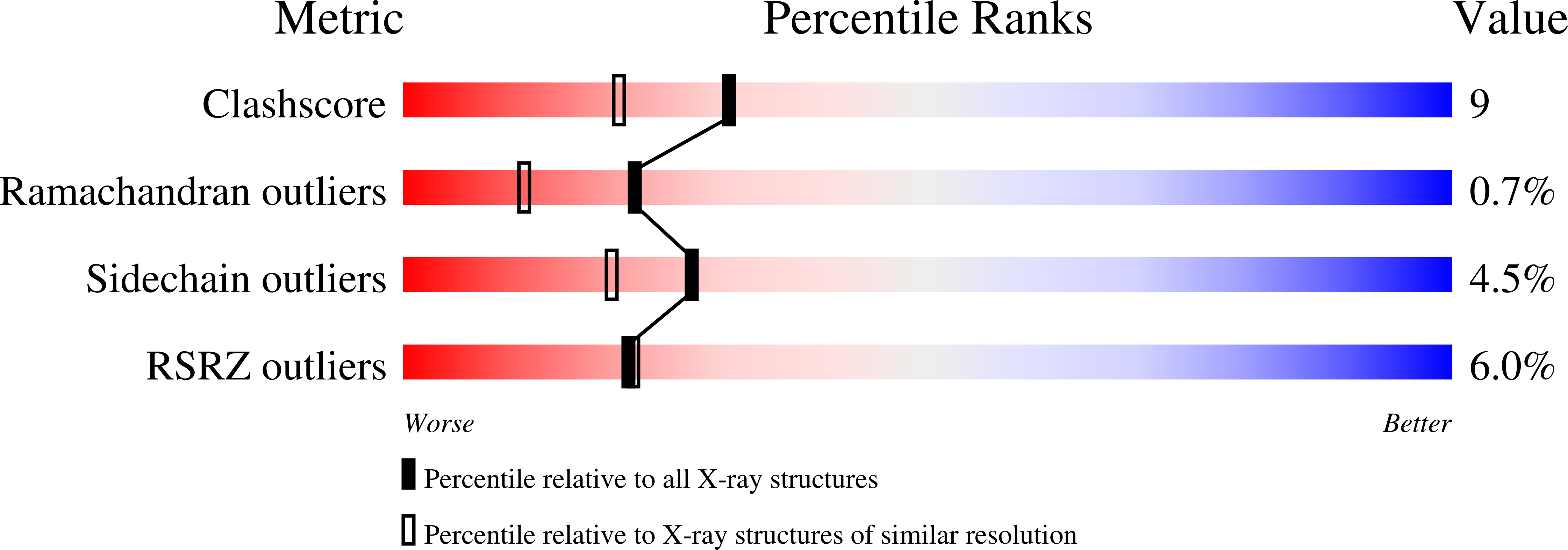
wwPDB Validation   3D Report Full Report


Ligand Structure Quality Assessment 


This is version 1.3 of the entry. See complete history


Literature

Ph Modulates the Quinone Position in the Photosynthetic Reaction Center from Rhodobacter Sphaeroides in the Neutral and Charge Separated States.

Koepke, J.Krammer, E.M.Klingen, A.R.Sebban, P.Ullmann, G.M.Fritzsch, G.

(2007) J Mol Biol 371: 396

  • DOI: https://doi.org/10.1016/j.jmb.2007.04.082
  • Primary Citation of Related Structures:  
    2J8C, 2J8D, 2UWS, 2UWT, 2UWU, 2UWV, 2UWW, 2UX3, 2UX4, 2UX5, 2UXJ, 2UXK, 2UXL, 2UXM

  • PubMed Abstract: 

    The structure of the photosynthetic reaction-center from Rhodobacter sphaeroides has been determined at four different pH values (6.5, 8.0, 9.0, 10.0) in the neutral and in charge separated states. At pH 8.0, in the neutral state, we obtain a resolution of 1.87 A, which is the best ever reported for the bacterial reaction center protein. Our crystallographic data confirm the existence of two different binding positions of the secondary quinone (QB). We observe a new orientation of QB in its distal position, which shows no ring-flip compared to the orientation in the proximal position. Datasets collected for the different pH values show a pH-dependence of the population of the proximal position. The new orientation of QB in the distal position and the pH-dependence could be confirmed by continuum electrostatics calculations. Our calculations are in agreement with the experimentally observed proton uptake upon charge separation. The high resolution of our crystallographic data allows us to identify new water molecules and external residues being involved in two previously described hydrogen bond proton channels. These extended proton-transfer pathways, ending at either of the two oxo-groups of QB in its proximal position, provide additional evidence that ring-flipping is not required for complete protonation of QB upon reduction.


  • Organizational Affiliation

    Max Planck Institute of Biophysics, Department of Molecular Membrane Biology, Max-von-Laue Strasse 3, D-60438 Frankfurt/Main, Germany. [email protected]


Macromolecules
Find similar proteins by:  (by identity cutoff)  |  3D Structure
Entity ID: 1
MoleculeChains Sequence LengthOrganismDetailsImage
REACTION CENTER PROTEIN H CHAINA [auth H]260Cereibacter sphaeroidesMutation(s): 0 
Membrane Entity: Yes 
UniProt
Find proteins for P0C0Y7 (Cereibacter sphaeroides)
Explore P0C0Y7 
Go to UniProtKB:  P0C0Y7
Entity Groups  
Sequence Clusters30% Identity50% Identity70% Identity90% Identity95% Identity100% Identity
UniProt GroupP0C0Y7
Sequence Annotations
Expand
  • Reference Sequence
Find similar proteins by:  (by identity cutoff)  |  3D Structure
Entity ID: 2
MoleculeChains Sequence LengthOrganismDetailsImage
REACTION CENTER PROTEIN L CHAINB [auth L]281Cereibacter sphaeroidesMutation(s): 0 
Membrane Entity: Yes 
UniProt
Find proteins for P0C0Y8 (Cereibacter sphaeroides)
Explore P0C0Y8 
Go to UniProtKB:  P0C0Y8
Entity Groups  
Sequence Clusters30% Identity50% Identity70% Identity90% Identity95% Identity100% Identity
UniProt GroupP0C0Y8
Sequence Annotations
Expand
  • Reference Sequence
Find similar proteins by:  (by identity cutoff)  |  3D Structure
Entity ID: 3
MoleculeChains Sequence LengthOrganismDetailsImage
REACTION CENTER PROTEIN M CHAINC [auth M]307Cereibacter sphaeroidesMutation(s): 0 
Membrane Entity: Yes 
UniProt
Find proteins for P0C0Y9 (Cereibacter sphaeroides)
Explore P0C0Y9 
Go to UniProtKB:  P0C0Y9
Entity Groups  
Sequence Clusters30% Identity50% Identity70% Identity90% Identity95% Identity100% Identity
UniProt GroupP0C0Y9
Sequence Annotations
Expand
  • Reference Sequence
Small Molecules
Ligands 10 Unique
IDChains Name / Formula / InChI Key2D Diagram3D Interactions
CDL
Query on CDL

Download Ideal Coordinates CCD File 
GA [auth M]CARDIOLIPIN
C81 H156 O17 P2
XVTUQDWPJJBEHJ-KZCWQMDCSA-L
BCL
Query on BCL

Download Ideal Coordinates CCD File 
H [auth L],
I [auth L],
U [auth M],
V [auth M]
BACTERIOCHLOROPHYLL A
C55 H74 Mg N4 O6
DSJXIQQMORJERS-AGGZHOMASA-M
BPH
Query on BPH

Download Ideal Coordinates CCD File 
DA [auth M],
M [auth L]
BACTERIOPHEOPHYTIN A
C55 H76 N4 O6
KWOZSBGNAHVCKG-SZQBJALDSA-N
U10
Query on U10

Download Ideal Coordinates CCD File 
EA [auth M],
N [auth L]
UBIQUINONE-10
C59 H90 O4
ACTIUHUUMQJHFO-UPTCCGCDSA-N
SPO
Query on SPO

Download Ideal Coordinates CCD File 
FA [auth M]SPHEROIDENE
C41 H60 O
FJOCMTHZSURUFA-KXCOHNEYSA-N
LDA
Query on LDA

Download Ideal Coordinates CCD File 
AA [auth M]
BA [auth M]
J [auth L]
K [auth L]
L
AA [auth M],
BA [auth M],
J [auth L],
K [auth L],
L,
W [auth M],
X [auth M],
Y [auth M],
Z [auth M]
LAURYL DIMETHYLAMINE-N-OXIDE
C14 H31 N O
SYELZBGXAIXKHU-UHFFFAOYSA-N
HTO
Query on HTO

Download Ideal Coordinates CCD File 
P [auth L],
Q [auth L]
HEPTANE-1,2,3-TRIOL
C7 H16 O3
HXYCHJFUBNTKQR-RNFRBKRXSA-N
PO4
Query on PO4

Download Ideal Coordinates CCD File 
O [auth L]PHOSPHATE ION
O4 P
NBIIXXVUZAFLBC-UHFFFAOYSA-K
GOL
Query on GOL

Download Ideal Coordinates CCD File 
D [auth H]
E [auth H]
F [auth H]
G [auth H]
R [auth L]
D [auth H],
E [auth H],
F [auth H],
G [auth H],
R [auth L],
S [auth L],
T [auth L]
GLYCEROL
C3 H8 O3
PEDCQBHIVMGVHV-UHFFFAOYSA-N
FE
Query on FE

Download Ideal Coordinates CCD File 
CA [auth M]FE (III) ION
Fe
VTLYFUHAOXGGBS-UHFFFAOYSA-N
Experimental Data & Validation

Experimental Data

  • Method: X-RAY DIFFRACTION
  • Resolution: 2.07 Å
  • R-Value Free: 0.218 
  • R-Value Work: 0.197 
  • R-Value Observed: 0.198 
  • Space Group: P 31 2 1
Unit Cell:
Length ( Å )Angle ( ˚ )
a = 140.01α = 90
b = 140.01β = 90
c = 184.76γ = 120
Software Package:
Software NamePurpose
REFMACrefinement
DENZOdata reduction
SCALEPACKdata scaling
CNSphasing

Structure Validation

View Full Validation Report


Ligand Structure Quality Assessment 


Entry History 

Deposition Data

Revision History  (Full details and data files)

  • Version 1.0: 2007-07-03
    Type: Initial release
  • Version 1.1: 2011-08-17
    Changes: Atomic model, Derived calculations, Non-polymer description, Other, Version format compliance
  • Version 1.2: 2019-07-24
    Changes: Data collection
  • Version 1.3: 2023-12-13
    Changes: Data collection, Database references, Derived calculations, Other, Refinement description