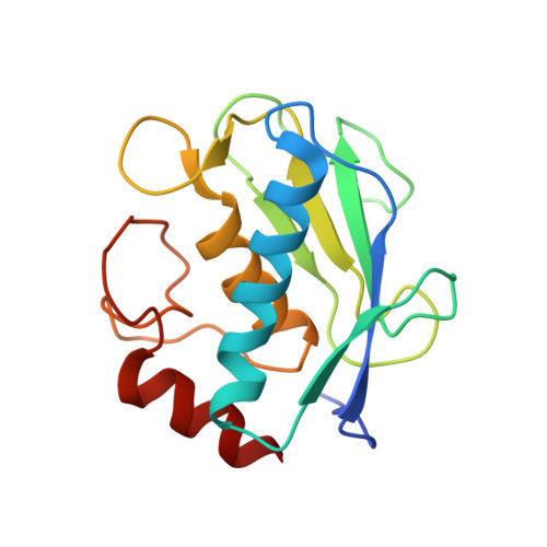
2OW2

MMP-9 active site mutant with difluoro butanoic acid inhibitor


Experimental Data Snapshot

  • Method: X-RAY DIFFRACTION
  • Resolution: 2.90 Å
  • R-Value Free: 0.285 
  • R-Value Work: 0.215 
  • R-Value Observed: 0.215 

Starting Model: experimental
View more details

wwPDB Validation   3D Report Full Report


Ligand Structure Quality Assessment 


This is version 1.5 of the entry. See complete history


Literature

Crystal Structures of MMP-9 Complexes with Five Inhibitors: Contribution of the Flexible Arg424 Side-chain to Selectivity.

Tochowicz, A.Maskos, K.Huber, R.Oltenfreiter, R.Dive, V.Yiotakis, A.Zanda, M.Bode, W.Goettig, P.

(2007) J Mol Biol 371: 989-1006

  • DOI: https://doi.org/10.1016/j.jmb.2007.05.068
  • Primary Citation of Related Structures:  
    2OVX, 2OVZ, 2OW0, 2OW1, 2OW2

  • PubMed Abstract: 

    Human matrix metalloproteinase 9 (MMP-9), also called gelatinase B, is particularly involved in inflammatory processes, bone remodelling and wound healing, but is also implicated in pathological processes such as rheumatoid arthritis, atherosclerosis, tumour growth, and metastasis. We have prepared the inactive E402Q mutant of the truncated catalytic domain of human MMP-9 and co-crystallized it with active site-directed synthetic inhibitors of different binding types. Here, we present the X-ray structures of five MMP-9 complexes with gelatinase-specific, tight binding inhibitors: a phosphinic acid (AM-409), a pyrimidine-2,4,6-trione (RO-206-0222), two carboxylate (An-1 and MJ-24), and a trifluoromethyl hydroxamic acid inhibitor (MS-560). These compounds bind by making a compromise between optimal coordination of the catalytic zinc, favourable hydrogen bond formation in the active-site cleft, and accommodation of their large hydrophobic P1' groups in the slightly flexible S1' cavity, which exhibits distinct rotational conformations of the Pro421 carbonyl group in each complex. In all these structures, the side-chain of Arg424 located at the bottom of the S1' cavity is not defined in the electron density beyond C(gamma), indicating its mobility. However, we suggest that the mobile Arg424 side-chain partially blocks the S1' cavity, which might explain the weaker binding of most inhibitors with a long P1' side-chain for MMP-9 compared with the closely related MMP-2 (gelatinase A), which exhibits a short threonine side-chain at the equivalent position. These novel structural details should facilitate the design of more selective MMP-9 inhibitors.


  • Organizational Affiliation

    Arbeitsgruppe Proteinaseforschung, Max-Planck-Institut für Biochemie, Am Klopferspitz 18, D-82152 Martinsried, Germany.


Macromolecules
Find similar proteins by:  (by identity cutoff)  |  3D Structure
Entity ID: 1
MoleculeChains Sequence LengthOrganismDetailsImage
Matrix metalloproteinase-9 (MMP-9) (92 kDa type IV collagenase) (92 kDa gelatinase) (Gelatinase B) (GELB)
A, B
159Homo sapiensMutation(s): 1 
Gene Names: MMP9CLG4B
EC: 3.4.24.35
UniProt & NIH Common Fund Data Resources
Find proteins for P14780 (Homo sapiens)
Explore P14780 
Go to UniProtKB:  P14780
PHAROS:  P14780
GTEx:  ENSG00000100985 
Entity Groups  
Sequence Clusters30% Identity50% Identity70% Identity90% Identity95% Identity100% Identity
UniProt GroupP14780
Sequence Annotations
Expand
  • Reference Sequence
Small Molecules
Ligands 4 Unique
IDChains Name / Formula / InChI Key2D Diagram3D Interactions
8MR
Query on 8MR

Download Ideal Coordinates CCD File 
J [auth A],
P [auth B]
(3R)-4,4-DIFLUORO-3-[(4-METHOXYPHENYL)SULFONYL]BUTANOIC ACID
C11 H12 F2 O5 S
OBQLOVWIRUXBAW-SECBINFHSA-N
ZN
Query on ZN

Download Ideal Coordinates CCD File 
C [auth A],
D [auth A],
K [auth B],
L [auth B]
ZINC ION
Zn
PTFCDOFLOPIGGS-UHFFFAOYSA-N
CA
Query on CA

Download Ideal Coordinates CCD File 
E [auth A]
F [auth A]
G [auth A]
M [auth B]
N [auth B]
E [auth A],
F [auth A],
G [auth A],
M [auth B],
N [auth B],
O [auth B]
CALCIUM ION
Ca
BHPQYMZQTOCNFJ-UHFFFAOYSA-N
CL
Query on CL

Download Ideal Coordinates CCD File 
H [auth A],
I [auth A]
CHLORIDE ION
Cl
VEXZGXHMUGYJMC-UHFFFAOYSA-M
Binding Affinity Annotations 
IDSourceBinding Affinity
8MR PDBBind:  2OW2 IC50: 6 (nM) from 1 assay(s)
Experimental Data & Validation

Experimental Data

  • Method: X-RAY DIFFRACTION
  • Resolution: 2.90 Å
  • R-Value Free: 0.285 
  • R-Value Work: 0.215 
  • R-Value Observed: 0.215 
  • Space Group: P 41 21 2
Unit Cell:
Length ( Å )Angle ( ˚ )
a = 55.42α = 90
b = 55.42β = 90
c = 260.793γ = 90
Software Package:
Software NamePurpose
CNSrefinement
DENZOdata reduction
SCALEPACKdata scaling
CNSphasing

Structure Validation

View Full Validation Report


Ligand Structure Quality Assessment 


Entry History 

Deposition Data

Revision History  (Full details and data files)

  • Version 1.0: 2007-06-19
    Type: Initial release
  • Version 1.1: 2008-05-01
    Changes: Version format compliance
  • Version 1.2: 2011-07-13
    Changes: Derived calculations, Version format compliance
  • Version 1.3: 2017-08-16
    Changes: Advisory, Source and taxonomy
  • Version 1.4: 2021-10-20
    Changes: Advisory, Database references, Derived calculations
  • Version 1.5: 2023-08-30
    Changes: Data collection, Refinement description