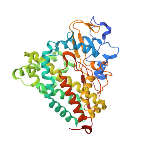
2QBN

Crystal structure of ferric G248V cytochrome P450cam


Experimental Data Snapshot

  • Method: X-RAY DIFFRACTION
  • Resolution: 1.75 Å
  • R-Value Free: 0.223 
  • R-Value Work: 0.196 

Starting Model: experimental
View more details

wwPDB Validation   3D Report Full Report


Ligand Structure Quality Assessment 


This is version 1.4 of the entry. See complete history


Literature

Alteration of P450 Distal Pocket Solvent Leads to Impaired Proton Delivery and Changes in Heme Geometry.

Makris, T.M.Koenig, K.V.Schlichting, I.Sligar, S.G.

(2007) Biochemistry 46: 14129-14140

  • DOI: https://doi.org/10.1021/bi7013695
  • Primary Citation of Related Structures:  
    2QBL, 2QBM, 2QBN, 2QBO

  • PubMed Abstract: 

    Distal pocket water molecules have been widely implicated in the delivery of protons required in O-O bond heterolysis in the P450 reaction cycle. Targeted dehydration of the cytochrome P450cam (CYP101) distal pocket through mutagenesis of a distal pocket glycine to either valine or threonine results in the alteration of spin state equilibria, and has dramatic consequences on the catalytic rate, coupling efficiency, and kinetic solvent isotope effect parameters, highlighting an important role of the active-site hydration level on P450 catalysis. Cryoradiolysis of the mutant CYP101 oxyferrous complexes further indicates a specific perturbation of proton-transfer events required for the transformation of ferric-peroxo to ferric-hydroperoxo states. Finally, crystallography of the 248Val and 248Thr mutants in both the ferric camphor bound resting state and ferric-cyano adducts shows both the alteration of hydrogen-bonding networks and the alteration of heme geometry parameters. Taken together, these results indicate that the distal pocket microenvironment governs the transformation of reactive heme-oxygen intermediates in P450 cytochromes.


  • Organizational Affiliation

    Department of Biochemistry, University of Illinois Urbana-Champaign, Urbana, Illinois 61801, USA.


Macromolecules
Find similar proteins by:  (by identity cutoff)  |  3D Structure
Entity ID: 1
MoleculeChains Sequence LengthOrganismDetailsImage
Cytochrome P450-cam421Pseudomonas putidaMutation(s): 1 
Gene Names: camCcyp101
EC: 1.14.15.1
UniProt
Find proteins for P00183 (Pseudomonas putida)
Explore P00183 
Go to UniProtKB:  P00183
Entity Groups  
Sequence Clusters30% Identity50% Identity70% Identity90% Identity95% Identity100% Identity
UniProt GroupP00183
Sequence Annotations
Expand
  • Reference Sequence
Experimental Data & Validation

Experimental Data

  • Method: X-RAY DIFFRACTION
  • Resolution: 1.75 Å
  • R-Value Free: 0.223 
  • R-Value Work: 0.196 
  • Space Group: P 43 21 2
Unit Cell:
Length ( Å )Angle ( ˚ )
a = 63.92α = 90
b = 63.92β = 90
c = 242.14γ = 90
Software Package:
Software NamePurpose
CNSrefinement
PDB_EXTRACTdata extraction
MAR345data collection
XDSdata reduction
XSCALEdata scaling
CNSphasing

Structure Validation

View Full Validation Report


Ligand Structure Quality Assessment 


Entry History 

Deposition Data

Revision History  (Full details and data files)

  • Version 1.0: 2007-12-25
    Type: Initial release
  • Version 1.1: 2011-07-13
    Changes: Version format compliance
  • Version 1.2: 2017-10-18
    Changes: Refinement description
  • Version 1.3: 2021-10-20
    Changes: Database references, Derived calculations
  • Version 1.4: 2023-08-30
    Changes: Data collection, Refinement description