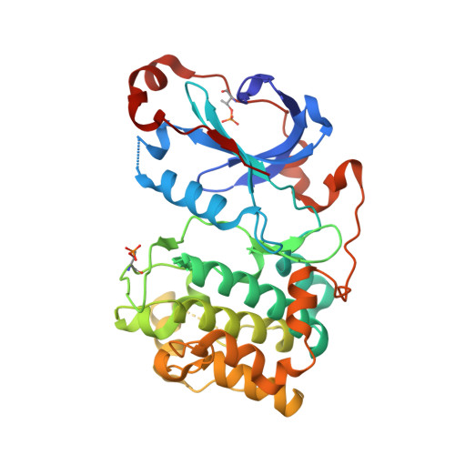
Structures of the PKC-iota kinase domain in its ATP-bound and apo forms reveal defined structures of residues 533-551 in the C-terminal tail and their roles in ATP binding
Takimura, T., Kamata, K., Fukasawa, K., Ohsawa, H., Komatani, H., Yoshizumi, T., Takahashi, I., Kotani, H., Iwasawa, Y.(2010) Acta Crystallogr D Biol Crystallogr 66: 577-583
- PubMed: 20445233
- DOI: https://doi.org/10.1107/S0907444910005639
- Primary Citation of Related Structures:
3A8W, 3A8X - PubMed Abstract:
Protein kinase C (PKC) plays an essential role in a wide range of cellular functions. Although crystal structures of the PKC-theta, PKC-iota and PKC-betaII kinase domains have previously been determined in complexes with small-molecule inhibitors, no structure of a PKC-substrate complex has been determined. In the previously determined PKC-iota complex, residues 533-551 in the C-terminal tail were disordered. In the present study, crystal structures of the PKC-iota kinase domain in its ATP-bound and apo forms were determined at 2.1 and 2.0 A resolution, respectively. In the ATP complex, the electron density of all of the C-terminal tail residues was well defined. In the structure, the side chain of Phe543 protrudes into the ATP-binding pocket to make van der Waals interactions with the adenine moiety of ATP; this is also observed in other AGC kinase structures such as binary and ternary substrate complexes of PKA and AKT. In addition to this interaction, the newly defined residues around the turn motif make multiple hydrogen bonds to glycine-rich-loop residues. These interactions reduce the flexibility of the glycine-rich loop, which is organized for ATP binding, and the resulting structure promotes an ATP conformation that is suitable for the subsequent phosphoryl transfer. In the case of the apo form, the structure and interaction mode of the C-terminal tail of PKC-iota are essentially identical to those of the ATP complex. These results indicate that the protein structure is pre-organized before substrate binding to PKC-iota, which is different from the case of the prototypical AGC-branch kinase PKA.
Organizational Affiliation:
Tsukuba Research Institute, Merck Research Laboratories, Banyu Pharmaceutical Co. Ltd, Okubo-3, Tsukuba, 300-2611 Ibaraki, Japan. [email protected]


