4JWS

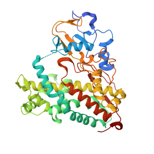
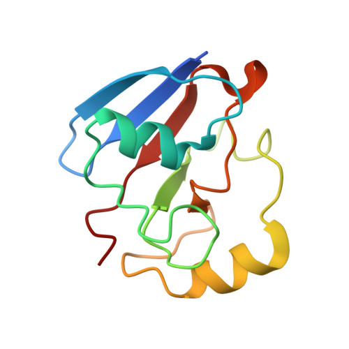
Crystal structure of Cytochrome P450cam-putidaredoxin complex


Experimental Data Snapshot

  • Method: X-RAY DIFFRACTION
  • Resolution: 2.15 Å
  • R-Value Free: 0.225 
  • R-Value Work: 0.175 
  • R-Value Observed: 0.178 

Starting Models: experimental
View more details

wwPDB Validation   3D Report Full Report


Ligand Structure Quality Assessment 


This is version 1.2 of the entry. See complete history


Literature

Structural basis for effector control and redox partner recognition in cytochrome P450.

Tripathi, S.Li, H.Poulos, T.L.

(2013) Science 340: 1227-1230

  • DOI: https://doi.org/10.1126/science.1235797
  • Primary Citation of Related Structures:  
    4JWS, 4JWU, 4JX1

  • PubMed Abstract: 

    Cytochromes P450 catalyze a variety of monooxygenase reactions that require electron transfer from redox partners. Although the structure of many P450s and a small handful of redox partners are known, there is very little structural information available on redox complexes, thus leaving a gap in our understanding on the control of P450-redox partner interactions. We have solved the crystal structure of oxidized and reduced P450cam complexed with its redox partner, putidaredoxin (Pdx), to 2.2 and 2.09 angstroms, respectively. It was anticipated that Pdx would favor closed substrate-bound P450cam, which differs substantially from the open conformer, but instead we found that Pdx favors the open state. These new structures indicate that the effector role of Pdx is to shift P450cam toward the open conformation, which enables the establishment of a water-mediated H-bonded network, which is required for proton-coupled electron transfer.


  • Organizational Affiliation

    Department of Molecular Biology and Biochemistry, University of California, Irvine, Irvine, CA 92697-3900, USA.


Macromolecules
Find similar proteins by:  (by identity cutoff)  |  3D Structure
Entity ID: 1
MoleculeChains Sequence LengthOrganismDetailsImage
Camphor 5-monooxygenase
A, B
415Pseudomonas putidaMutation(s): 6 
Gene Names: camCcyp101
EC: 1.14.15.1
UniProt
Find proteins for P00183 (Pseudomonas putida)
Explore P00183 
Go to UniProtKB:  P00183
Entity Groups  
Sequence Clusters30% Identity50% Identity70% Identity90% Identity95% Identity100% Identity
UniProt GroupP00183
Sequence Annotations
Expand
  • Reference Sequence
Find similar proteins by:  (by identity cutoff)  |  3D Structure
Entity ID: 2
MoleculeChains Sequence LengthOrganismDetailsImage
Putidaredoxin
C, D
112Pseudomonas putidaMutation(s): 0 
Gene Names: camB
UniProt
Find proteins for P00259 (Pseudomonas putida)
Explore P00259 
Go to UniProtKB:  P00259
Entity Groups  
Sequence Clusters30% Identity50% Identity70% Identity90% Identity95% Identity100% Identity
UniProt GroupP00259
Sequence Annotations
Expand
  • Reference Sequence
Small Molecules
Ligands 4 Unique
IDChains Name / Formula / InChI Key2D Diagram3D Interactions
HEM
Query on HEM

Download Ideal Coordinates CCD File 
E [auth A],
H [auth B]
PROTOPORPHYRIN IX CONTAINING FE
C34 H32 Fe N4 O4
KABFMIBPWCXCRK-RGGAHWMASA-L
1N0
Query on 1N0

Download Ideal Coordinates CCD File 
G [auth A],
J [auth B]
1,1'-hexane-1,6-diyldipyrrolidine-2,5-dione
C14 H20 N2 O4
PBFKSBAPGGMKKJ-UHFFFAOYSA-N
FES
Query on FES

Download Ideal Coordinates CCD File 
K [auth C],
L [auth D]
FE2/S2 (INORGANIC) CLUSTER
Fe2 S2
NIXDOXVAJZFRNF-UHFFFAOYSA-N
CA
Query on CA

Download Ideal Coordinates CCD File 
F [auth A],
I [auth B]
CALCIUM ION
Ca
BHPQYMZQTOCNFJ-UHFFFAOYSA-N
Experimental Data & Validation

Experimental Data

  • Method: X-RAY DIFFRACTION
  • Resolution: 2.15 Å
  • R-Value Free: 0.225 
  • R-Value Work: 0.175 
  • R-Value Observed: 0.178 
  • Space Group: P 1 21 1
Unit Cell:
Length ( Å )Angle ( ˚ )
a = 57.59α = 90
b = 106.72β = 107.91
c = 88.7γ = 90
Software Package:
Software NamePurpose
BOSdata collection
PHASERphasing
PHENIXrefinement
MOSFLMdata reduction
SCALAdata scaling

Structure Validation

View Full Validation Report


Ligand Structure Quality Assessment 


Entry History 

Deposition Data

Revision History  (Full details and data files)

  • Version 1.0: 2013-06-19
    Type: Initial release
  • Version 1.1: 2023-09-20
    Changes: Data collection, Database references, Derived calculations, Refinement description
  • Version 1.2: 2024-10-16
    Changes: Structure summary