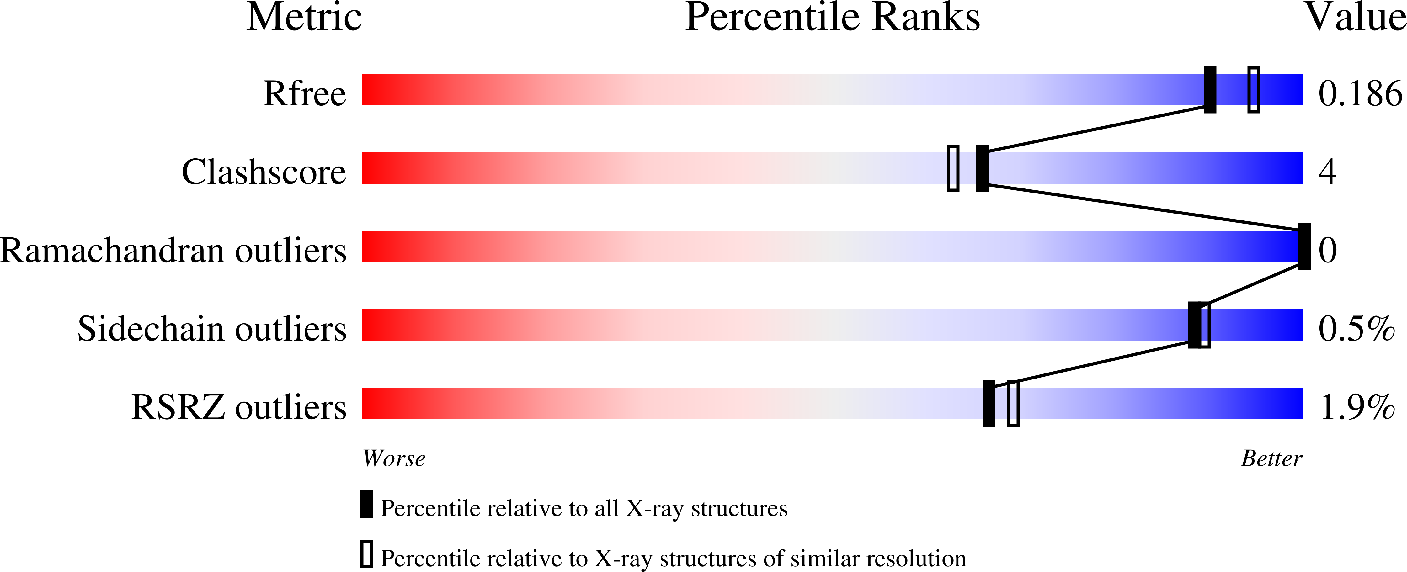
Identification of a new hormone-binding site on the surface of thyroid hormone receptor.
Souza, P.C., Puhl, A.C., Martinez, L., Aparicio, R., Nascimento, A.S., Figueira, A.C., Nguyen, P., Webb, P., Skaf, M.S., Polikarpov, I.(2014) Mol Endocrinol 28: 534-545
- PubMed: 24552590
- DOI: https://doi.org/10.1210/me.2013-1359
- Primary Citation of Related Structures:
4LNW, 4LNX - PubMed Abstract:
Thyroid hormone receptors (TRs) are members of the nuclear receptor superfamily of ligand-activated transcription factors involved in cell differentiation, growth, and homeostasis. Although X-ray structures of many nuclear receptor ligand-binding domains (LBDs) reveal that the ligand binds within the hydrophobic core of the ligand-binding pocket, a few studies suggest the possibility of ligands binding to other sites. Here, we report a new x-ray crystallographic structure of TR-LBD that shows a second binding site for T3 and T4 located between H9, H10, and H11 of the TRα LBD surface. Statistical multiple sequence analysis, site-directed mutagenesis, and cell transactivation assays indicate that residues of the second binding site could be important for the TR function. We also conducted molecular dynamics simulations to investigate ligand mobility and ligand-protein interaction for T3 and T4 bound to this new TR surface-binding site. Extensive molecular dynamics simulations designed to compute ligand-protein dissociation constant indicate that the binding affinities to this surface site are of the order of the plasma and intracellular concentrations of the thyroid hormones, suggesting that ligands may bind to this new binding site under physiological conditions. Therefore, the second binding site could be useful as a new target site for drug design and could modulate selectively TR functions.
Organizational Affiliation:
Institute of Chemistry (P.C.T.S., L.M., R.A., M.S.S.), State University of Campinas-UNICAMP, Campinas, Sao Paulo, Brazil; Institute of Physics of São Carlos (A.C.P., A.S.N., P.W., I.P.), University of São Paulo-USP, São Carlos, Sao Paulo, Brazil; National Laboratory of Biosciences (A.C.M.F.), CNPEM, Campinas, Sao Paulo, Brazil; University of California Medical Center (P.N.), Diabetes Center, San Francisco, California; and Genomic Medicine (P.W.), Houston Methodist Research Institute, Houston, Texas.


