4Q0D

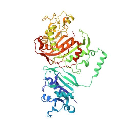
Crystal structure of TS-DHFR from Cryptosporidium hominis in complex with NADPH, FdUMP, methotrexate and 2-amino-4-oxo-4,7-dihydro-pyrrolo[2,3-d]pyrimidine-methyl-phenyl-L-glutamic acid.


Experimental Data Snapshot

  • Method: X-RAY DIFFRACTION
  • Resolution: 3.45 Å
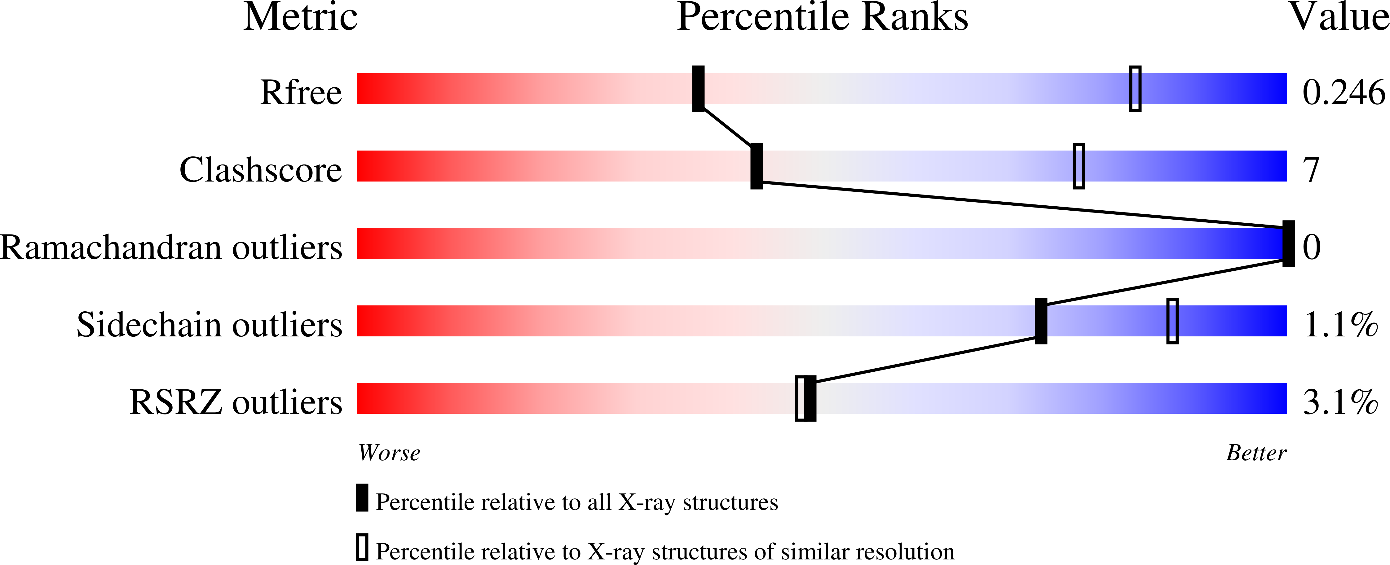
  • R-Value Free: 0.242 
  • R-Value Work: 0.217 
  • R-Value Observed: 0.218 

Starting Model: experimental
View more details

wwPDB Validation   3D Report Full Report


Ligand Structure Quality Assessment 


This is version 1.2 of the entry. See complete history


Literature

Structural studies provide clues for analog design of specific inhibitors of Cryptosporidium hominis thymidylate synthase-dihydrofolate reductase.

Kumar, V.P.Cisneros, J.A.Frey, K.M.Castellanos-Gonzalez, A.Wang, Y.Gangjee, A.White, A.C.Jorgensen, W.L.Anderson, K.S.

(2014) Bioorg Med Chem Lett 24: 4158-4161

  • DOI: https://doi.org/10.1016/j.bmcl.2014.07.049
  • Primary Citation of Related Structures:  
    4Q0D, 4Q0E

  • PubMed Abstract: 

    Cryptosporidium is the causative agent of a gastrointestinal disease, cryptosporidiosis, which is often fatal in immunocompromised individuals and children. Thymidylate synthase (TS) and dihydrofolate reductase (DHFR) are essential enzymes in the folate biosynthesis pathway and are well established as drug targets in cancer, bacterial infections, and malaria. Cryptosporidium hominis has a bifunctional thymidylate synthase and dihydrofolate reductase enzyme, compared to separate enzymes in the host. We evaluated lead compound 1 from a novel series of antifolates, 2-amino-4-oxo-5-substituted pyrrolo[2,3-d]pyrimidines as an inhibitor of Cryptosporidium hominis thymidylate synthase with selectivity over the human enzyme. Complementing the enzyme inhibition compound 1 also has anti-cryptosporidial activity in cell culture. A crystal structure with compound 1 bound to the TS active site is discussed in terms of several van der Waals, hydrophobic and hydrogen bond interactions with the protein residues and the substrate analog 5-fluorodeoxyuridine monophosphate (TS), cofactor NADPH and inhibitor methotrexate (DHFR). Another crystal structure in complex with compound 1 bound in both the TS and DHFR active sites is also reported here. The crystal structures provide clues for analog design and for the design of ChTS-DHFR specific inhibitors.


  • Organizational Affiliation

    Department of Pharmacology, Yale University School of Medicine, 333 Cedar Street, New Haven, CT 06520, USA.


Macromolecules
Find similar proteins by:  (by identity cutoff)  |  3D Structure
Entity ID: 1
MoleculeChains Sequence LengthOrganismDetailsImage
Bifunctional dihydrofolate reductase-thymidylate synthaseA [auth E],
B [auth A],
C [auth B],
D [auth C],
E [auth D]
521Cryptosporidium hominisMutation(s): 0 
Gene Names: Chro.40506TS-DHFR
EC: 2.1.1.45 (PDB Primary Data), 1.5.1.3 (PDB Primary Data)
Entity Groups  
Sequence Clusters30% Identity50% Identity70% Identity90% Identity95% Identity100% Identity
Sequence Annotations
Expand
  • Reference Sequence
Small Molecules
Ligands 4 Unique
IDChains Name / Formula / InChI Key2D Diagram3D Interactions
NDP
Query on NDP

Download Ideal Coordinates CCD File 
F [auth E],
J [auth A],
N [auth B],
R [auth C],
V [auth D]
NADPH DIHYDRO-NICOTINAMIDE-ADENINE-DINUCLEOTIDE PHOSPHATE
C21 H30 N7 O17 P3
ACFIXJIJDZMPPO-NNYOXOHSSA-N
MTX
Query on MTX

Download Ideal Coordinates CCD File 
I [auth E],
M [auth A],
Q [auth B],
U [auth C],
Y [auth D]
METHOTREXATE
C20 H22 N8 O5
FBOZXECLQNJBKD-ZDUSSCGKSA-N
2XB
Query on 2XB

Download Ideal Coordinates CCD File 
H [auth E],
L [auth A],
P [auth B],
T [auth C],
X [auth D]
N-{4-[(2-amino-4-hydroxy-7H-pyrrolo[2,3-d]pyrimidin-5-yl)methyl]benzoyl}-L-glutamic acid
C19 H19 N5 O6
VDHBMZRBPMTLGE-LBPRGKRZSA-N
UFP
Query on UFP

Download Ideal Coordinates CCD File 
G [auth E],
K [auth A],
O [auth B],
S [auth C],
W [auth D]
5-FLUORO-2'-DEOXYURIDINE-5'-MONOPHOSPHATE
C9 H12 F N2 O8 P
HFEKDTCAMMOLQP-RRKCRQDMSA-N
Experimental Data & Validation

Experimental Data

  • Method: X-RAY DIFFRACTION
  • Resolution: 3.45 Å
  • R-Value Free: 0.242 
  • R-Value Work: 0.217 
  • R-Value Observed: 0.218 
  • Space Group: C 1 2 1
Unit Cell:
Length ( Å )Angle ( ˚ )
a = 213.611α = 90
b = 115.031β = 93.92
c = 217.643γ = 90
Software Package:
Software NamePurpose
DENZOdata reduction
SCALEPACKdata scaling
PHENIXrefinement
PDB_EXTRACTdata extraction
CBASSdata collection
HKL-2000data reduction
HKL-2000data scaling
PHASERphasing

Structure Validation

View Full Validation Report


Ligand Structure Quality Assessment 


Entry History 

Deposition Data

Revision History  (Full details and data files)

  • Version 1.0: 2014-10-15
    Type: Initial release
  • Version 1.1: 2017-11-22
    Changes: Refinement description
  • Version 1.2: 2023-09-20
    Changes: Data collection, Database references, Derived calculations, Refinement description