5LV1

2.12 A resolution structure of PtxB from Prochlorococcus marinus (MIT 9301) in complex with phosphite


Experimental Data Snapshot

  • Method: X-RAY DIFFRACTION
  • Resolution: 2.12 Å
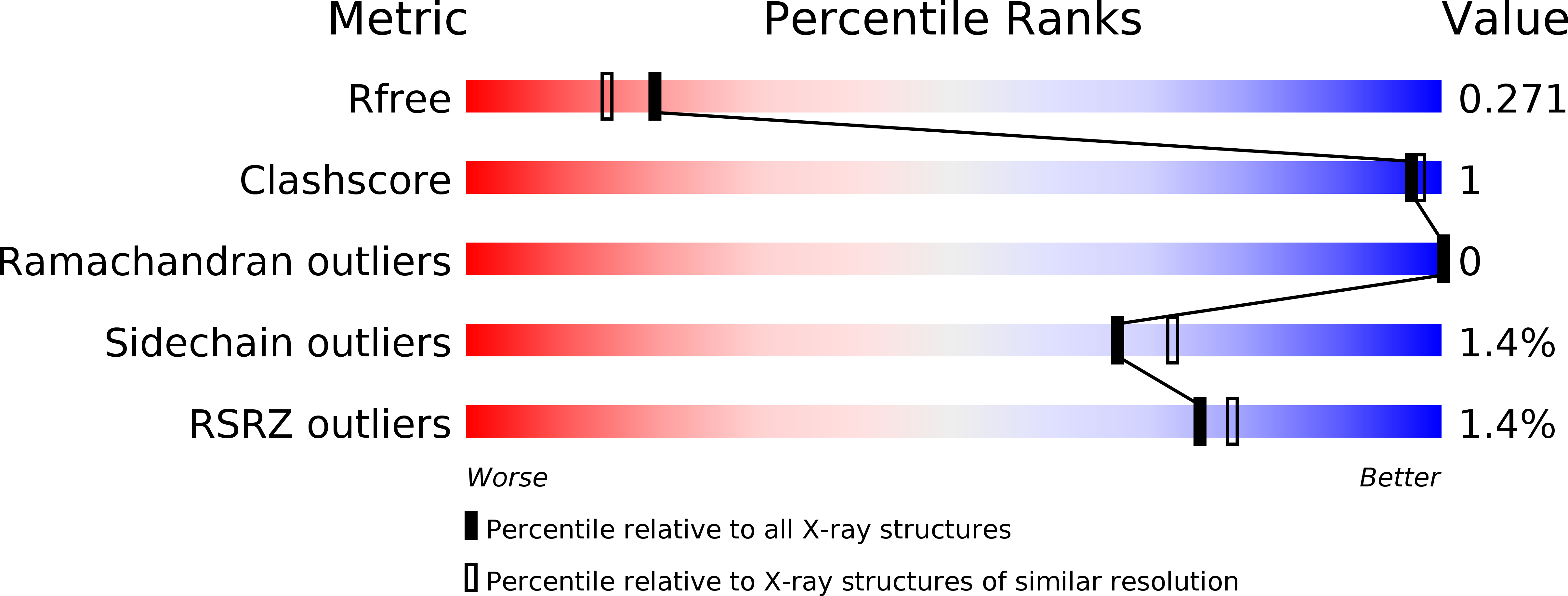
  • R-Value Free: 0.264 
  • R-Value Work: 0.218 
  • R-Value Observed: 0.220 

Starting Model: experimental
View more details

wwPDB Validation   3D Report Full Report


This is version 1.2 of the entry. See complete history


Literature

The molecular basis of phosphite and hypophosphite recognition by ABC-transporters.

Bisson, C.Adams, N.B.P.Stevenson, B.Brindley, A.A.Polyviou, D.Bibby, T.S.Baker, P.J.Hunter, C.N.Hitchcock, A.

(2017) Nat Commun 8: 1746-1746

  • DOI: https://doi.org/10.1038/s41467-017-01226-8
  • Primary Citation of Related Structures:  
    5JVB, 5LQ1, 5LQ5, 5LQ8, 5LV1, 5ME4, 5O2J, 5O2K, 5O37

  • PubMed Abstract: 

    Inorganic phosphate is the major bioavailable form of the essential nutrient phosphorus. However, the concentration of phosphate in most natural habitats is low enough to limit microbial growth. Under phosphate-depleted conditions some bacteria utilise phosphite and hypophosphite as alternative sources of phosphorus, but the molecular basis of reduced phosphorus acquisition from the environment is not fully understood. Here, we present crystal structures and ligand binding affinities of periplasmic binding proteins from bacterial phosphite and hypophosphite ATP-binding cassette transporters. We reveal that phosphite and hypophosphite specificity results from a combination of steric selection and the presence of a P-H…π interaction between the ligand and a conserved aromatic residue in the ligand-binding pocket. The characterisation of high affinity and specific transporters has implications for the marine phosphorus redox cycle, and might aid the use of phosphite as an alternative phosphorus source in biotechnological, industrial and agricultural applications.


  • Organizational Affiliation

    Department of Molecular Biology and Biotechnology, University of Sheffield, Firth Court, Western Bank, Sheffield, S10 2TN, UK.


Macromolecules
Find similar proteins by:  (by identity cutoff)  |  3D Structure
Entity ID: 1
MoleculeChains Sequence LengthOrganismDetailsImage
PtxBA,
B [auth C],
C [auth B]
280Prochlorococcus marinus str. MIT 9301Mutation(s): 0 
Gene Names: P9301_12511
UniProt
Find proteins for A3PDP9 (Prochlorococcus marinus (strain MIT 9301))
Explore A3PDP9 
Go to UniProtKB:  A3PDP9
Entity Groups  
Sequence Clusters30% Identity50% Identity70% Identity90% Identity95% Identity100% Identity
UniProt GroupA3PDP9
Sequence Annotations
Expand
  • Reference Sequence
Experimental Data & Validation

Experimental Data

  • Method: X-RAY DIFFRACTION
  • Resolution: 2.12 Å
  • R-Value Free: 0.264 
  • R-Value Work: 0.218 
  • R-Value Observed: 0.220 
  • Space Group: P 31 2 1
Unit Cell:
Length ( Å )Angle ( ˚ )
a = 152.175α = 90
b = 152.175β = 90
c = 67.915γ = 120
Software Package:
Software NamePurpose
REFMACrefinement
XDSdata reduction
XSCALEdata scaling
PHASERphasing

Structure Validation

View Full Validation Report


Entry History & Funding Information

Deposition Data


Funding OrganizationLocationGrant Number
Biotechnology and Biological Sciences Research CouncilUnited KingdomBB/M000265/1
Biotechnology and Biological Sciences Research CouncilUnited KingdomBB/M011305/1
European Research CouncilUnited Kingdom338895
NERC/A. G. Leventis FoundationUnited KingdomPhD studentship

Revision History  (Full details and data files)

  • Version 1.0: 2017-12-06
    Type: Initial release
  • Version 1.1: 2018-01-24
    Changes: Source and taxonomy
  • Version 1.2: 2024-01-17
    Changes: Data collection, Database references, Refinement description