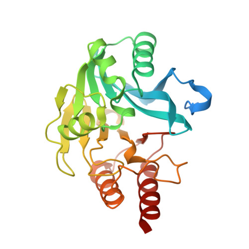
5N55

mono-Zinc VIM-5 metallo-beta-lactamase in complex with (1-chloro-4-hydroxyisoquinoline-3-carbonyl)-L-tryptophan (Compound 2)


Experimental Data Snapshot

  • Method: X-RAY DIFFRACTION
  • Resolution: 1.99 Å
  • R-Value Free: 0.253 
  • R-Value Work: 0.191 
  • R-Value Observed: 0.194 

Starting Model: experimental
View more details

wwPDB Validation   3D Report Full Report


Ligand Structure Quality Assessment 


This is version 1.2 of the entry. See complete history


Literature

Crystallographic analyses of isoquinoline complexes reveal a new mode of metallo-beta-lactamase inhibition.

Li, G.B.Brem, J.Lesniak, R.Abboud, M.I.Lohans, C.T.Clifton, I.J.Yang, S.Y.Jimenez-Castellanos, J.C.Avison, M.B.Spencer, J.McDonough, M.A.Schofield, C.J.

(2017) Chem Commun (Camb) 53: 5806-5809

  • DOI: https://doi.org/10.1039/c7cc02394d
  • Primary Citation of Related Structures:  
    5N4S, 5N4T, 5N55, 5N58, 5NAI

  • PubMed Abstract: 

    Crystallographic analyses of the VIM-5 metallo-β-lactamase (MBL) with isoquinoline inhibitors reveal non zinc ion binding modes. Comparison with other MBL-inhibitor structures directed addition of a zinc-binding thiol enabling identification of potent B1 MBL inhibitors. The inhibitors potentiate meropenem activity against clinical isolates harboring MBLs.


  • Organizational Affiliation

    Department of Chemistry, University of Oxford, 12 Mansfield Road, Oxford, OX1 3TA, UK. [email protected] [email protected].


Macromolecules
Find similar proteins by:  (by identity cutoff)  |  3D Structure
Entity ID: 1
MoleculeChains Sequence LengthOrganismDetailsImage
Class B metallo-beta-lactamase
A, B, C
266Klebsiella pneumoniaeMutation(s): 0 
Gene Names: blaVIM-5
EC: 3.5.2.6
UniProt
Find proteins for Q8GKX2 (Klebsiella pneumoniae)
Explore Q8GKX2 
Go to UniProtKB:  Q8GKX2
Entity Groups  
Sequence Clusters30% Identity50% Identity70% Identity90% Identity95% Identity100% Identity
UniProt GroupQ8GKX2
Sequence Annotations
Expand
  • Reference Sequence
Experimental Data & Validation

Experimental Data

  • Method: X-RAY DIFFRACTION
  • Resolution: 1.99 Å
  • R-Value Free: 0.253 
  • R-Value Work: 0.191 
  • R-Value Observed: 0.194 
  • Space Group: F 2 2 2
Unit Cell:
Length ( Å )Angle ( ˚ )
a = 66.219α = 90
b = 155.191β = 90
c = 267.733γ = 90
Software Package:
Software NamePurpose
SCALEPACKdata scaling
PHASERphasing
PHENIXrefinement
PDB_EXTRACTdata extraction
HKL-3000data reduction

Structure Validation

View Full Validation Report


Ligand Structure Quality Assessment 


Entry History 

Deposition Data

Revision History  (Full details and data files)

  • Version 1.0: 2017-05-17
    Type: Initial release
  • Version 1.1: 2017-06-07
    Changes: Database references
  • Version 1.2: 2024-01-31
    Changes: Data collection, Database references, Derived calculations, Refinement description