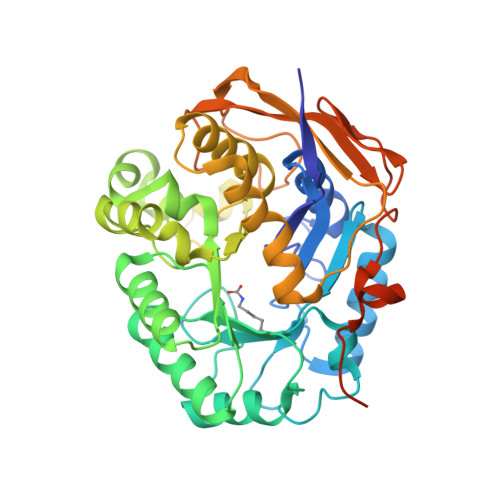
6HFR

Human dihydroorotase mutant F1563Y co-crystallized with carbamoyl aspartate at pH 7.0


Experimental Data Snapshot

  • Method: X-RAY DIFFRACTION
  • Resolution: 1.30 Å
  • R-Value Free: 0.144 
  • R-Value Work: 0.126 
  • R-Value Observed: 0.127 

Starting Model: experimental
View more details

wwPDB Validation   3D Report Full Report


Ligand Structure Quality Assessment 


This is version 1.2 of the entry. See complete history


Literature

Characterization of the catalytic flexible loop in the dihydroorotase domain of the human multi-enzymatic protein CAD.

Del Cano-Ochoa, F.Grande-Garcia, A.Reverte-Lopez, M.D'Abramo, M.Ramon-Maiques, S.

(2018) J Biol Chem 293: 18903-18913

  • DOI: https://doi.org/10.1074/jbc.RA118.005494
  • Primary Citation of Related Structures:  
    6HFD, 6HFE, 6HFF, 6HFH, 6HFI, 6HFJ, 6HFK, 6HFL, 6HFN, 6HFP, 6HFQ, 6HFR, 6HFS, 6HFU, 6HG1, 6HG2, 6HG3

  • PubMed Abstract: 

    The dihydroorotase (DHOase) domain of the multifunctional protein carbamoyl-phosphate synthetase 2, aspartate transcarbamoylase, and dihydroorotase (CAD) catalyzes the third step in the de novo biosynthesis of pyrimidine nucleotides in animals. The crystal structure of the DHOase domain of human CAD (huDHOase) revealed that, despite evolutionary divergence, its active site components are highly conserved with those in bacterial DHOases, encoded as monofunctional enzymes. An important element for catalysis, conserved from Escherichia coli to humans, is a flexible loop that closes as a lid over the active site. Here, we combined mutagenic, structural, biochemical, and molecular dynamics analyses to characterize the function of the flexible loop in the activity of CAD's DHOase domain. A huDHOase chimera bearing the E. coli DHOase flexible loop was inactive, suggesting the presence of distinctive elements in the flexible loop of huDHOase that cannot be replaced by the bacterial sequence. We pinpointed Phe-1563, a residue absolutely conserved at the tip of the flexible loop in CAD's DHOase domain, as a critical element for the conformational equilibrium between the two catalytic states of the protein. Substitutions of Phe-1563 with Ala, Leu, or Thr prevented the closure of the flexible loop and inactivated the protein, whereas substitution with Tyr enhanced the interactions of the loop in the closed position and reduced fluctuations and the reaction rate. Our results confirm the importance of the flexible loop in CAD's DHOase domain and explain the key role of Phe-1563 in configuring the active site and in promoting substrate strain and catalysis.


  • Organizational Affiliation

    From the Department of Genome Dynamics and Function, Centro de Biología Molecular Severo Ochoa (CSIC-UAM), Madrid 28049, Spain.


Macromolecules
Find similar proteins by:  (by identity cutoff)  |  3D Structure
Entity ID: 1
MoleculeChains Sequence LengthOrganismDetailsImage
CAD protein393Homo sapiensMutation(s): 1 
Gene Names: CAD
EC: 6.3.5.5 (PDB Primary Data), 2.1.3.2 (PDB Primary Data), 3.5.2.3 (PDB Primary Data), 6.3.4.16 (UniProt), 3.5.1.2 (UniProt)
UniProt & NIH Common Fund Data Resources
Find proteins for P27708 (Homo sapiens)
Explore P27708 
Go to UniProtKB:  P27708
PHAROS:  P27708
GTEx:  ENSG00000084774 
Entity Groups  
Sequence Clusters30% Identity50% Identity70% Identity90% Identity95% Identity100% Identity
UniProt GroupP27708
Sequence Annotations
Expand
  • Reference Sequence
Small Molecules
Ligands 3 Unique
IDChains Name / Formula / InChI Key2D Diagram3D Interactions
NCD
Query on NCD

Download Ideal Coordinates CCD File 
E [auth A]N-CARBAMOYL-L-ASPARTATE
C5 H8 N2 O5
HLKXYZVTANABHZ-REOHCLBHSA-N
ZN
Query on ZN

Download Ideal Coordinates CCD File 
B [auth A],
C [auth A],
D [auth A],
I [auth A]
ZINC ION
Zn
PTFCDOFLOPIGGS-UHFFFAOYSA-N
FMT
Query on FMT

Download Ideal Coordinates CCD File 
F [auth A],
G [auth A],
H [auth A]
FORMIC ACID
C H2 O2
BDAGIHXWWSANSR-UHFFFAOYSA-N
Modified Residues  1 Unique
IDChains TypeFormula2D DiagramParent
KCX
Query on KCX
A
L-PEPTIDE LINKINGC7 H14 N2 O4LYS
Experimental Data & Validation

Experimental Data

  • Method: X-RAY DIFFRACTION
  • Resolution: 1.30 Å
  • R-Value Free: 0.144 
  • R-Value Work: 0.126 
  • R-Value Observed: 0.127 
  • Space Group: C 2 2 21
Unit Cell:
Length ( Å )Angle ( ˚ )
a = 81.863α = 90
b = 159.088β = 90
c = 60.989γ = 90
Software Package:
Software NamePurpose
PHENIXrefinement
XDSdata reduction
Aimlessdata scaling
PHASERphasing

Structure Validation

View Full Validation Report


Ligand Structure Quality Assessment 


Entry History & Funding Information

Deposition Data


Funding OrganizationLocationGrant Number
Spanish Ministry of Economy and CompetitivenessSpainBFU2016-80570-R

Revision History  (Full details and data files)

  • Version 1.0: 2018-10-24
    Type: Initial release
  • Version 1.1: 2018-12-19
    Changes: Data collection, Database references
  • Version 1.2: 2024-01-17
    Changes: Data collection, Database references, Derived calculations, Refinement description