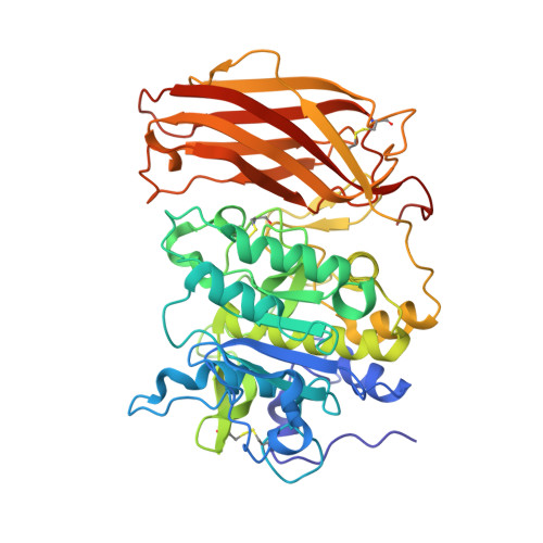
6HZC

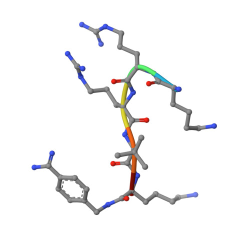
X-ray structure of furin in complex with the cyclic inhibitor c[glutaryl-BVK-Lys-Arg-Arg-Tle-Lys]-4-Amba


Experimental Data Snapshot

  • Method: X-RAY DIFFRACTION
  • Resolution: 1.90 Å
  • R-Value Free: 0.183 
  • R-Value Work: 0.164 
  • R-Value Observed: 0.165 

Starting Model: experimental
View more details

wwPDB Validation   3D Report Full Report


This is version 1.4 of the entry. See complete history


Literature

Design, Synthesis, and Characterization of Macrocyclic Inhibitors of the Proprotein Convertase Furin.

Van Lam van, T.Ivanova, T.Hardes, K.Heindl, M.R.Morty, R.E.Bottcher-Friebertshauser, E.Lindberg, I.Than, M.E.Dahms, S.O.Steinmetzer, T.

(2019) ChemMedChem 14: 673-685

  • DOI: https://doi.org/10.1002/cmdc.201800807
  • Primary Citation of Related Structures:  
    6HLB, 6HLD, 6HLE, 6HZA, 6HZB, 6HZC, 6HZD

  • PubMed Abstract: 

    The activation of viral glycoproteins by the host protease furin is an essential step in the replication of numerous pathogenic viruses. Thus, effective inhibitors of furin could serve as broad-spectrum antiviral drugs. A crystal structure of an inhibitory hexapeptide derivative in complex with furin served as template for the rational design of various types of new cyclic inhibitors. Most of the prepared derivatives are relatively potent furin inhibitors with inhibition constants in the low nanomolar or even sub-nanomolar range. For seven derivatives the crystal structures in complex with furin could be determined. In three complexes, electron density was found for the entire inhibitor. In the other cases the structures could be determined only for the P6/P5-P1 segments, which directly interact with furin. The cyclic derivatives together with two non-cyclic reference compounds were tested as inhibitors of the proteolytic activation and replication of respiratory syncytial virus in cells. Significant antiviral activity was found for both linear reference inhibitors, whereas a negligible efficacy was determined for the cyclic derivatives.


  • Organizational Affiliation

    Institute of Pharmaceutical Chemistry, Philipps University, Marbacher Weg 6, 35032, Marburg, Germany.


Macromolecules
Find similar proteins by:  (by identity cutoff)  |  3D Structure
Entity ID: 1
MoleculeChains Sequence LengthOrganismDetailsImage
Furin482Homo sapiensMutation(s): 0 
Gene Names: FURINFURPACEPCSK3
EC: 3.4.21.75
UniProt & NIH Common Fund Data Resources
Find proteins for P09958 (Homo sapiens)
Explore P09958 
Go to UniProtKB:  P09958
PHAROS:  P09958
GTEx:  ENSG00000140564 
Entity Groups  
Sequence Clusters30% Identity50% Identity70% Identity90% Identity95% Identity100% Identity
UniProt GroupP09958
Sequence Annotations
Expand
  • Reference Sequence

Find similar proteins by:  Sequence   |   3D Structure  

Entity ID: 2
MoleculeChains Sequence LengthOrganismDetailsImage
LYS-ARG-ARG-TBG-LYS-00S6synthetic constructMutation(s): 0 
Sequence Annotations
Expand
  • Reference Sequence
Small Molecules
Ligands 7 Unique
IDChains Name / Formula / InChI Key2D Diagram3D Interactions
BVK
Query on BVK

Download Ideal Coordinates CCD File 
N [auth B]2-[4-(aminomethyl)phenyl]ethanoic acid
C9 H11 N O2
HAAUVXXFRQXTTQ-UHFFFAOYSA-N
PTD
Query on PTD

Download Ideal Coordinates CCD File 
M [auth B]PENTANEDIAL
C5 H8 O2
SXRSQZLOMIGNAQ-UHFFFAOYSA-N
PO4
Query on PO4

Download Ideal Coordinates CCD File 
J [auth A],
K [auth A]
PHOSPHATE ION
O4 P
NBIIXXVUZAFLBC-UHFFFAOYSA-K
DMS
Query on DMS

Download Ideal Coordinates CCD File 
L [auth A]DIMETHYL SULFOXIDE
C2 H6 O S
IAZDPXIOMUYVGZ-UHFFFAOYSA-N
CA
Query on CA

Download Ideal Coordinates CCD File 
C [auth A],
D [auth A],
E [auth A]
CALCIUM ION
Ca
BHPQYMZQTOCNFJ-UHFFFAOYSA-N
CL
Query on CL

Download Ideal Coordinates CCD File 
I [auth A]CHLORIDE ION
Cl
VEXZGXHMUGYJMC-UHFFFAOYSA-M
NA
Query on NA

Download Ideal Coordinates CCD File 
F [auth A],
G [auth A],
H [auth A]
SODIUM ION
Na
FKNQFGJONOIPTF-UHFFFAOYSA-N
Modified Residues  1 Unique
IDChains TypeFormula2D DiagramParent
TBG
Query on TBG
B
L-PEPTIDE LINKINGC6 H13 N O2VAL
Experimental Data & Validation

Experimental Data

  • Method: X-RAY DIFFRACTION
  • Resolution: 1.90 Å
  • R-Value Free: 0.183 
  • R-Value Work: 0.164 
  • R-Value Observed: 0.165 
  • Space Group: P 65 2 2
Unit Cell:
Length ( Å )Angle ( ˚ )
a = 131.689α = 90
b = 131.689β = 90
c = 155.339γ = 120
Software Package:
Software NamePurpose
PHENIXrefinement
XDSdata reduction
XDSdata scaling
PHENIXphasing

Structure Validation

View Full Validation Report


Entry History 

Deposition Data

  • Released Date: 2019-02-06 
  • Deposition Author(s): Dahms, S.O.

Revision History  (Full details and data files)

  • Version 1.0: 2019-02-06
    Type: Initial release
  • Version 1.1: 2019-02-27
    Changes: Data collection, Database references
  • Version 1.2: 2019-04-03
    Changes: Data collection, Database references
  • Version 1.3: 2024-01-24
    Changes: Advisory, Data collection, Database references, Derived calculations, Refinement description
  • Version 1.4: 2024-11-20
    Changes: Structure summary