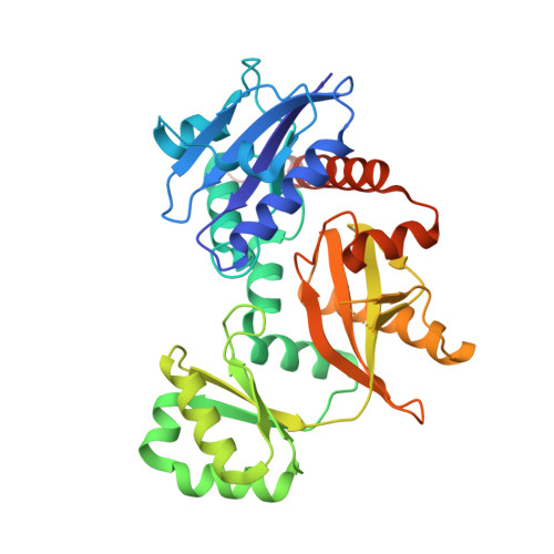
7U9K

Staphylococcus aureus D-alanine-D-alanine ligase in complex with ATP, D-ala-D-ala, Mg2+ and K+


Experimental Data Snapshot

  • Method: X-RAY DIFFRACTION
  • Resolution: 2.00 Å
  • R-Value Free: 0.277 
  • R-Value Work: 0.239 
  • R-Value Observed: 0.241 

Starting Model: experimental
View more details

wwPDB Validation   3D Report Full Report


Ligand Structure Quality Assessment 


This is version 1.3 of the entry. See complete history


Literature

Structure-guided design and synthesis of ATP-competitive N-acyl-substituted sulfamide d-alanine-d-alanine ligase inhibitors.

Becker, R.Pederick, J.L.Dawes, E.G.Bruning, J.B.Abell, A.D.

(2023) Bioorg Med Chem 96: 117509-117509

  • DOI: https://doi.org/10.1016/j.bmc.2023.117509
  • Primary Citation of Related Structures:  
    7U9K

  • PubMed Abstract: 

    d-Alanine-d-alanine ligase (Ddl) catalyses the ATP-dependent formation of d-Ala-d-Ala, a critical component in bacterial cell wall biosynthesis and is a validated target for new antimicrobial agents. Here, we describe the structure-guided design, synthesis, and evaluation of ATP-competitive N-acyl-substituted sulfamides 27-36, 42, 46, 47 as inhibitors of Staphylococcus aureus Ddl (SaDdl). A crystal structure of SaDdl complexed with ATP and d-Ala-d-Ala (PDB: 7U9K) identified ATP-mimetic 8 as an initial scaffold for further inhibitor design. Evaluation of 8 in SaDdl enzyme inhibition assays revealed the ability to reduce enzyme activity to 72 ± 8 % (IC 50  = 1.6 mM). The sulfamide linker of 8 was extended with 2-(4-methoxyphenyl)ethanol to give 29, to investigate further interactions with the d-Ala pocket of SaDdl, as predicted by molecular docking. This compound reduced enzyme activity to 89 ± 1 %, with replacement of the 4-methoxyphenyl group in 29 with alternative phenyl substituents (27, 28, 31-33, 35, 36) failing to significantly improve on this (80-89 % remaining enzyme activity). Exchanging these phenyl substituents with selected heterocycles (42, 46, 47) did improve activity, with the most active compound (42) reducing SaDdl activity to 70 ± 1 % (IC 50  = 1.7 mM), which compares favourably to the FDA-approved inhibitor d-cycloserine (DCS) (IC 50  = 0.1 mM). To the best of our knowledge, this is the first reported study of bisubstrate SaDdl inhibitors.


  • Organizational Affiliation

    Department of Chemistry, School of Physics, Chemistry and Earth Sciences, University of Adelaide, Adelaide, South Australia 5005, Australia; Institute for Photonics and Advanced Sensing, (IPAS), School of Biological Sciences, The University of Adelaide, Adelaide, South Australia 5005, Australia; Centre for Nanoscale BioPhotonics (CNBP), University of Adelaide, Adelaide, South Australia 5005, Australia.


Macromolecules
Find similar proteins by:  (by identity cutoff)  |  3D Structure
Entity ID: 1
MoleculeChains Sequence LengthOrganismDetailsImage
D-alanine--D-alanine ligase
A, B
364Staphylococcus aureus subsp. aureus NCTC 8325Mutation(s): 0 
Gene Names: ddlSAOUHSC_02318
EC: 6.3.2.4
UniProt
Find proteins for Q2FWH3 (Staphylococcus aureus (strain NCTC 8325 / PS 47))
Explore Q2FWH3 
Go to UniProtKB:  Q2FWH3
Entity Groups  
Sequence Clusters30% Identity50% Identity70% Identity90% Identity95% Identity100% Identity
UniProt GroupQ2FWH3
Sequence Annotations
Expand
  • Reference Sequence

Find similar proteins by:  Sequence   |   3D Structure  

Entity ID: 2
MoleculeChains Sequence LengthOrganismDetailsImage
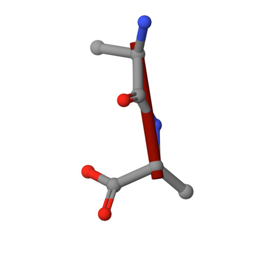
DAL-DALC [auth F]2Staphylococcus aureus subsp. aureus NCTC 8325Mutation(s): 0 
Sequence Annotations
Expand
  • Reference Sequence
Small Molecules
Ligands 4 Unique
IDChains Name / Formula / InChI Key2D Diagram3D Interactions
ATP (Subject of Investigation/LOI)
Query on ATP

Download Ideal Coordinates CCD File 
K [auth B]ADENOSINE-5'-TRIPHOSPHATE
C10 H16 N5 O13 P3
ZKHQWZAMYRWXGA-KQYNXXCUSA-N
ADP
Query on ADP

Download Ideal Coordinates CCD File 
F [auth A]ADENOSINE-5'-DIPHOSPHATE
C10 H15 N5 O10 P2
XTWYTFMLZFPYCI-KQYNXXCUSA-N
K (Subject of Investigation/LOI)
Query on K

Download Ideal Coordinates CCD File 
D [auth A],
G [auth B],
H [auth B],
I [auth B]
POTASSIUM ION
K
NPYPAHLBTDXSSS-UHFFFAOYSA-N
MG (Subject of Investigation/LOI)
Query on MG

Download Ideal Coordinates CCD File 
E [auth A],
J [auth B]
MAGNESIUM ION
Mg
JLVVSXFLKOJNIY-UHFFFAOYSA-N
Experimental Data & Validation

Experimental Data

  • Method: X-RAY DIFFRACTION
  • Resolution: 2.00 Å
  • R-Value Free: 0.277 
  • R-Value Work: 0.239 
  • R-Value Observed: 0.241 
  • Space Group: P 1 21 1
Unit Cell:
Length ( Å )Angle ( ˚ )
a = 52.447α = 90
b = 70.058β = 102.58
c = 91.598γ = 90
Software Package:
Software NamePurpose
PHENIXrefinement
XDSdata reduction
Aimlessdata scaling
PHASERphasing
PDB_EXTRACTdata extraction

Structure Validation

View Full Validation Report


Ligand Structure Quality Assessment 


Entry History & Funding Information

Deposition Data


Funding OrganizationLocationGrant Number
Not funded--

Revision History  (Full details and data files)

  • Version 1.0: 2023-03-15
    Type: Initial release
  • Version 1.1: 2023-10-25
    Changes: Data collection, Refinement description
  • Version 1.2: 2024-01-31
    Changes: Database references
  • Version 1.3: 2024-11-13
    Changes: Structure summary