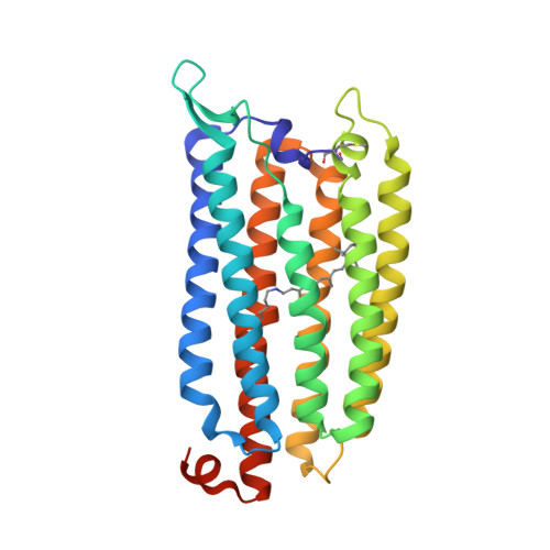
8QQZ

Cryo-EM structure of the light-driven sodium pump ErNaR in the pentameric form at pH 8.0


Experimental Data Snapshot

  • Method: ELECTRON MICROSCOPY
  • Resolution: 2.63 Å
  • Aggregation State: PARTICLE 
  • Reconstruction Method: SINGLE PARTICLE 

wwPDB Validation   3D Report Full Report


This is version 1.0 of the entry. See complete history


Literature

A subgroup of light-driven sodium pumps with an additional Schiff base counterion.

Podoliak, E.Lamm, G.H.U.Marin, E.Schellbach, A.V.Fedotov, D.A.Stetsenko, A.Asido, M.Maliar, N.Bourenkov, G.Balandin, T.Baeken, C.Astashkin, R.Schneider, T.R.Bateman, A.Wachtveitl, J.Schapiro, I.Busskamp, V.Guskov, A.Gordeliy, V.Alekseev, A.Kovalev, K.

(2024) Nat Commun 15: 3119-3119

  • DOI: https://doi.org/10.1038/s41467-024-47469-0
  • Primary Citation of Related Structures:  
    8QLE, 8QLF, 8QQZ, 8QR0

  • PubMed Abstract: 

    Light-driven sodium pumps (NaRs) are unique ion-transporting microbial rhodopsins. The major group of NaRs is characterized by an NDQ motif and has two aspartic acid residues in the central region essential for sodium transport. Here we identify a subgroup of the NDQ rhodopsins bearing an additional glutamic acid residue in the close vicinity to the retinal Schiff base. We thoroughly characterize a member of this subgroup, namely the protein ErNaR from Erythrobacter sp. HL-111 and show that the additional glutamic acid results in almost complete loss of pH sensitivity for sodium-pumping activity, which is in contrast to previously studied NaRs. ErNaR is capable of transporting sodium efficiently even at acidic pH levels. X-ray crystallography and single particle cryo-electron microscopy reveal that the additional glutamic acid residue mediates the connection between the other two Schiff base counterions and strongly interacts with the aspartic acid of the characteristic NDQ motif. Hence, it reduces its pKa. Our findings shed light on a subgroup of NaRs and might serve as a basis for their rational optimization for optogenetics.


  • Organizational Affiliation

    Department of Ophthalmology, University Hospital Bonn, Medical Faculty, Bonn, Germany.


Macromolecules
Find similar proteins by:  (by identity cutoff)  |  3D Structure
Entity ID: 1
MoleculeChains Sequence LengthOrganismDetailsImage
Bacteriorhodopsin-like protein
A, B, C, D, E
283ErythrobacterMutation(s): 0 
Gene Names: SAMN04515621_2824
Membrane Entity: Yes 
UniProt
Find proteins for A0A1H1XA63 (Erythrobacter sp. HL-111)
Explore A0A1H1XA63 
Go to UniProtKB:  A0A1H1XA63
Entity Groups  
Sequence Clusters30% Identity50% Identity70% Identity90% Identity95% Identity100% Identity
UniProt GroupA0A1H1XA63
Sequence Annotations
Expand
  • Reference Sequence
Small Molecules
Ligands 2 Unique
IDChains Name / Formula / InChI Key2D Diagram3D Interactions
LMT
Query on LMT

Download Ideal Coordinates CCD File 
BA [auth B]
CA [auth B]
IB [auth E]
LA [auth C]
MA [auth C]
BA [auth B],
CA [auth B],
IB [auth E],
LA [auth C],
MA [auth C],
P [auth A],
Q [auth A],
R [auth A],
YA [auth D],
ZA [auth D]
DODECYL-BETA-D-MALTOSIDE
C24 H46 O11
NLEBIOOXCVAHBD-QKMCSOCLSA-N
LFA
Query on LFA

Download Ideal Coordinates CCD File 
AA [auth B]
AB [auth E]
BB [auth E]
CB [auth E]
DA [auth C]
AA [auth B],
AB [auth E],
BB [auth E],
CB [auth E],
DA [auth C],
DB [auth E],
EA [auth C],
EB [auth E],
F [auth A],
FA [auth C],
FB [auth E],
G [auth A],
GA [auth C],
GB [auth E],
H [auth A],
HA [auth C],
HB [auth E],
I [auth A],
IA [auth C],
J [auth A],
JA [auth C],
K [auth A],
KA [auth C],
L [auth A],
M [auth A],
N [auth A],
NA [auth D],
O [auth A],
OA [auth D],
PA [auth D],
QA [auth D],
RA [auth D],
S [auth B],
SA [auth D],
T [auth B],
TA [auth D],
U [auth B],
UA [auth D],
V [auth B],
VA [auth D],
W [auth B],
WA [auth D],
X [auth B],
XA [auth D],
Y [auth B],
Z [auth B]
EICOSANE
C20 H42
CBFCDTFDPHXCNY-UHFFFAOYSA-N
Modified Residues  2 Unique
IDChains TypeFormula2D DiagramParent
FME
Query on FME
A, B, C, D, E
L-PEPTIDE LINKINGC6 H11 N O3 SMET
LYR
Query on LYR
A, B, C, D, E
L-PEPTIDE LINKINGC26 H42 N2 O2LYS
Experimental Data & Validation

Experimental Data

  • Method: ELECTRON MICROSCOPY
  • Resolution: 2.63 Å
  • Aggregation State: PARTICLE 
  • Reconstruction Method: SINGLE PARTICLE 
EM Software:
TaskSoftware PackageVersion
RECONSTRUCTIONcryoSPARC4.0.2

Structure Validation

View Full Validation Report


Entry History & Funding Information

Deposition Data


Funding OrganizationLocationGrant Number
EIPOD fellowship under Marie Sklodowska-Curie Actions COFUNDGermany847543

Revision History  (Full details and data files)

  • Version 1.0: 2024-04-24
    Type: Initial release