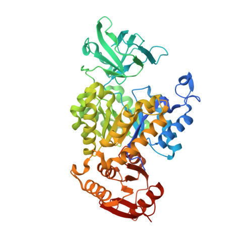
8XW8

Crystal structure of Streptococcus pneumoniae pyruvate kinase in complex with oxalate and fructose 1,6-bisphosphate and GDP


Experimental Data Snapshot

  • Method: X-RAY DIFFRACTION
  • Resolution: 2.00 Å
  • R-Value Free: 0.234 
  • R-Value Work: 0.198 
  • R-Value Observed: 0.200 

Starting Model: experimental
View more details

wwPDB Validation   3D Report Full Report


Ligand Structure Quality Assessment 


This is version 1.1 of the entry. See complete history


Literature

Structural Basis of Nucleotide Selectivity in Pyruvate Kinase.

Taguchi, A.Nakashima, R.Nishino, K.

(2024) J Mol Biol 436: 168708-168708

  • DOI: https://doi.org/10.1016/j.jmb.2024.168708
  • Primary Citation of Related Structures:  
    8XW6, 8XW7, 8XW8, 8XW9, 8ZLY

  • PubMed Abstract: 

    Nucleoside triphosphates are indispensable in numerous biological processes, with enzymes involved in their biogenesis playing pivotal roles in cell proliferation. Pyruvate kinase (PYK), commonly regarded as the terminal glycolytic enzyme that generates ATP in tandem with pyruvate, is also capable of synthesizing a wide range of nucleoside triphosphates from their diphosphate precursors. Despite their substrate promiscuity, some PYKs show preference towards specific nucleotides, suggesting an underlying mechanism for differentiating nucleotide bases. However, the thorough characterization of this mechanism has been hindered by the paucity of nucleotide-bound PYK structures. Here, we present crystal structures of Streptococcus pneumoniae PYK in complex with four different nucleotides. These structures facilitate direct comparison of the protein-nucleotide interactions and offer structural insights into its pronounced selectivity for GTP synthesis. Notably, this selectivity is dependent on a sequence motif in the nucleotide recognition site that is widely present among prokaryotic PYKs, particularly in Firmicutes species. We show that pneumococcal cell growth is significantly impaired when expressing a PYK variant with compromised GTP and UTP synthesis activity, underscoring the importance of PYK in maintaining nucleotide homeostasis. Our findings collectively advance our understanding of PYK biochemistry and prokaryotic metabolism.


  • Organizational Affiliation

    SANKEN, Osaka University, Ibaraki, Osaka 567-0047, Japan; Graduate School of Pharmaceutical Sciences, Osaka University, Suita, Osaka 565-0871, Japan. Electronic address: [email protected].


Macromolecules
Find similar proteins by:  (by identity cutoff)  |  3D Structure
Entity ID: 1
MoleculeChains Sequence LengthOrganismDetailsImage
Pyruvate kinase
A, B, C, D
521Streptococcus pneumoniae R6Mutation(s): 0 
Gene Names: pykF
EC: 2.7.1.40
UniProt
Find proteins for Q8DQ84 (Streptococcus pneumoniae (strain ATCC BAA-255 / R6))
Explore Q8DQ84 
Go to UniProtKB:  Q8DQ84
Entity Groups  
Sequence Clusters30% Identity50% Identity70% Identity90% Identity95% Identity100% Identity
UniProt GroupQ8DQ84
Sequence Annotations
Expand
  • Reference Sequence
Small Molecules
Ligands 6 Unique
IDChains Name / Formula / InChI Key2D Diagram3D Interactions
GDP
Query on GDP

Download Ideal Coordinates CCD File 
FA [auth D],
I [auth A],
Q [auth B],
Y [auth C]
GUANOSINE-5'-DIPHOSPHATE
C10 H15 N5 O11 P2
QGWNDRXFNXRZMB-UUOKFMHZSA-N
FBP
Query on FBP

Download Ideal Coordinates CCD File 
EA [auth D],
H [auth A],
P [auth B],
X [auth C]
1,6-di-O-phosphono-beta-D-fructofuranose
C6 H14 O12 P2
RNBGYGVWRKECFJ-ARQDHWQXSA-N
GOL
Query on GOL

Download Ideal Coordinates CCD File 
AA [auth C]
HA [auth D]
K [auth A]
L [auth A]
S [auth B]
AA [auth C],
HA [auth D],
K [auth A],
L [auth A],
S [auth B],
T [auth B]
GLYCEROL
C3 H8 O3
PEDCQBHIVMGVHV-UHFFFAOYSA-N
OXL
Query on OXL

Download Ideal Coordinates CCD File 
GA [auth D],
J [auth A],
R [auth B],
Z [auth C]
OXALATE ION
C2 O4
MUBZPKHOEPUJKR-UHFFFAOYSA-L
K
Query on K

Download Ideal Coordinates CCD File 
DA [auth D],
G [auth A],
O [auth B],
W [auth C]
POTASSIUM ION
K
NPYPAHLBTDXSSS-UHFFFAOYSA-N
MG
Query on MG

Download Ideal Coordinates CCD File 
BA [auth D]
CA [auth D]
E [auth A]
F [auth A]
M [auth B]
BA [auth D],
CA [auth D],
E [auth A],
F [auth A],
M [auth B],
N [auth B],
U [auth C],
V [auth C]
MAGNESIUM ION
Mg
JLVVSXFLKOJNIY-UHFFFAOYSA-N
Experimental Data & Validation

Experimental Data

  • Method: X-RAY DIFFRACTION
  • Resolution: 2.00 Å
  • R-Value Free: 0.234 
  • R-Value Work: 0.198 
  • R-Value Observed: 0.200 
  • Space Group: C 1 2 1
Unit Cell:
Length ( Å )Angle ( ˚ )
a = 123.335α = 90
b = 256.08β = 90.04
c = 88.575γ = 90
Software Package:
Software NamePurpose
REFMACrefinement
XDSdata scaling
XDSdata reduction
MOLREPphasing

Structure Validation

View Full Validation Report


Ligand Structure Quality Assessment 


Entry History & Funding Information

Deposition Data


Funding OrganizationLocationGrant Number
Japan Society for the Promotion of Science (JSPS)Japan21K20759

Revision History  (Full details and data files)

  • Version 1.0: 2024-07-31
    Type: Initial release
  • Version 1.1: 2024-08-07
    Changes: Database references