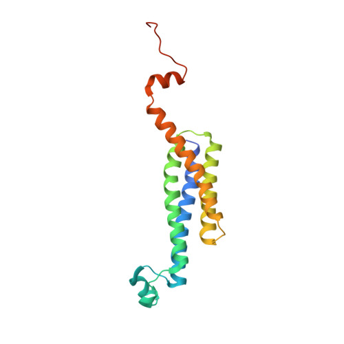
4BPM

Crystal structure of a human integral membrane enzyme


Experimental Data Snapshot

  • Method: X-RAY DIFFRACTION
  • Resolution: 2.08 Å
  • R-Value Free: 0.218 
  • R-Value Work: 0.200 
  • R-Value Observed: 0.201 

wwPDB Validation   3D Report Full Report


Ligand Structure Quality Assessment 


This is version 1.5 of the entry. See complete history


Literature

Crystallizing Membrane Proteins in the Lipidic Mesophase. Experience with Human Prostaglandin E2 Synthase 1 and an Evolving Strategy.

Li, D.Howe, N.Dukkipati, A.Shah, S.T.A.Bax, B.D.Edge, C.Bridges, A.Hardwicke, P.Singh, O.M.P.Giblin, G.Pautsch, A.Pfau, R.Schnapp, G.Wang, M.Olieric, V.Caffrey, M.

(2014) Cryst Growth Des 14: 2034

  • DOI: https://doi.org/10.1021/cg500157x
  • Primary Citation of Related Structures:  
    4BPM

  • PubMed Abstract: 

    The lipidic mesophase or in meso method for crystallizing membrane proteins has several high profile targets to its credit and is growing in popularity. Despite its success, the method is in its infancy as far as rational crystallogenesis is concerned. Consequently, significant time, effort, and resources are still required to generate structure-grade crystals, especially with a new target type. Therefore, a need exists for crystallogenesis protocols that are effective with a broad range of membrane protein types. Recently, a strategy for crystallizing a prokaryotic α-helical membrane protein, diacylglycerol kinase (DgkA), by the in meso method was reported (Cryst. Growth. Des.2013, 14, 2846-2857). Here, we describe its application to the human α-helical microsomal prostaglandin E2 synthase 1 (mPGES1). While the DgkA strategy proved useful, significant modifications were needed to generate structure-quality crystals of this important therapeutic target. These included protein engineering, using an additive phospholipid in the hosting mesophase, performing multiple rounds of salt screening, and carrying out trials at 4 °C in the presence of a tight binding ligand. The crystallization strategy detailed here should prove useful for generating structures of other integral membrane proteins by the in meso method.


  • Organizational Affiliation

    Membrane Structural and Functional Biology Group, School of Medicine and School of Biochemistry and Immunology, Trinity College Dublin, Dublin 2, Ireland.


Macromolecules
Find similar proteins by:  (by identity cutoff)  |  3D Structure
Entity ID: 1
MoleculeChains Sequence LengthOrganismDetailsImage
PROSTAGLANDIN E SYNTHASE, FUSION PEPTIDE178Homo sapienssynthetic construct
This entity is chimeric
Mutation(s): 1 
EC: 5.3.99.3 (PDB Primary Data), 1.11.1 (UniProt), 2.5.1 (UniProt), 4.4.1.20 (UniProt), 2.5.1.18 (UniProt)
Membrane Entity: Yes 
UniProt & NIH Common Fund Data Resources
Find proteins for O14684 (Homo sapiens)
Explore O14684 
Go to UniProtKB:  O14684
PHAROS:  O14684
GTEx:  ENSG00000148344 
Find proteins for Q16873 (Homo sapiens)
Explore Q16873 
Go to UniProtKB:  Q16873
PHAROS:  Q16873
Entity Groups  
Sequence Clusters30% Identity50% Identity70% Identity90% Identity95% Identity100% Identity
UniProt GroupsO14684Q16873
Sequence Annotations
Expand
  • Reference Sequence
Small Molecules
Ligands 2 Unique
IDChains Name / Formula / InChI Key2D Diagram3D Interactions
LVJ
Query on LVJ

Download Ideal Coordinates CCD File 
C [auth A]2-[[2,6-bis(chloranyl)-3-[(2,2-dimethylpropanoylamino)methyl]phenyl]amino]-1-methyl-6-(2-methyl-2-oxidanyl-propoxy)-N-[2,2,2-tris(fluoranyl)ethyl]benzimidazole-5-carboxamide
C27 H32 Cl2 F3 N5 O4
CHFOASGROVVRSK-UHFFFAOYSA-N
GSH
Query on GSH

Download Ideal Coordinates CCD File 
B [auth A]GLUTATHIONE
C10 H17 N3 O6 S
RWSXRVCMGQZWBV-WDSKDSINSA-N
Experimental Data & Validation

Experimental Data

  • Method: X-RAY DIFFRACTION
  • Resolution: 2.08 Å
  • R-Value Free: 0.218 
  • R-Value Work: 0.200 
  • R-Value Observed: 0.201 
  • Space Group: H 3 2
Unit Cell:
Length ( Å )Angle ( ˚ )
a = 86.4α = 90
b = 86.4β = 90
c = 181.12γ = 120
Software Package:
Software NamePurpose
PHENIXrefinement
XDSdata reduction
XSCALEdata scaling
SHELXphasing

Structure Validation

View Full Validation Report


Ligand Structure Quality Assessment 


Entry History 

Deposition Data

Revision History  (Full details and data files)

  • Version 1.0: 2014-04-16
    Type: Initial release
  • Version 1.1: 2014-05-14
    Changes: Database references
  • Version 1.2: 2017-03-15
    Changes: Source and taxonomy
  • Version 1.3: 2019-03-06
    Changes: Data collection, Experimental preparation
  • Version 1.4: 2019-04-03
    Changes: Data collection, Experimental preparation
  • Version 1.5: 2024-05-08
    Changes: Data collection, Database references, Derived calculations, Other